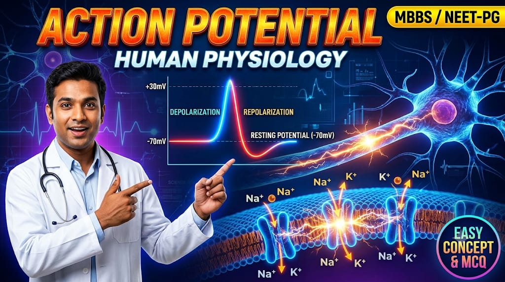
In human physiology ,the action potential is the fundamental electrical signals through which excitable cells -neurons and muscle fibers communicate, contract and transmit rapid signals across the body.
An action potential is a rapid, brief sequence of changes in the membrane potential that occurs after excitation.
A stimulation that causes a rapid sequence of changes in the resting membrane potential of an excitable cell is known as an action potential.
Do you know types of excitable cells?
Yes, there are two types of excitable cellsà Muscle cells and Nerve cells.
RMP (Resting Membrane Potential) is between -70 and -100 millivolts.
1. The Resting Membrane Potential
When stimulated, the resting membrane potential changes due to a disturbance in ionic equilibrium across the receptor zone of the cell membrane.
Action potential generation occurs in Phases.
The Phases are:
The latent period is the time taken by the impulses to travel along the axon from the site of stimulation to the recording electrode. It is expressed in milliseconds.
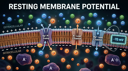
The Resting Membrane Potential is between -70 and -100 millivolts. This is because the interior of the cell membrane is negative relative to the exterior. Potassium ion permeability is greater than that of sodium ions.
The size and duration of an action potential vary and depend on the nature of the tissue. For example, in nerves, it is no more than a few milliseconds, while in cardiac muscle, it is 200 to 300 milliseconds.
Outer positive and inner negative near the cell membrane.
Point of stimulus
The point of stimulus is the point or place on the cell membrane where the stimulus is applied.
When a stimulus is applied, it produces a brief, irregular baseline deflection due to current leakage from the stimulating electrode to the recording electrode. This is the point of stimulus. At the point of stimulation, a slight decrease in the RMP is due to an increase in K+ influx, which is restored by K+ efflux.
2. Latent Period
The latent period depends on-
Speed of conduction of impulses in the axon-character of the axon. And
Distance between the stimulus and recording electrode.
We can calculate the speed of conduction in a nerve by applying a stimulus and recording the time to response, and measuring the distance.
The stimulus artifact is followed by an iso potential interval –without any obvious change -which ends with the next potential change.
Depolarization
Depolarization is the first indication the approaching impulse. Depolarization is the change from a negative to a positive membrane potential.
At the point of stimulation, a slight decrease in the RMP is due to increased K+ influx, which is counteracted by K+ efflux.
When stimulation persists sodium leaky channel opens, sodium enters the cell, and the membrane potential rises up to +7 m V. But potassium efflux increases through the leaky channels and restore membrane potential. If another stimulus reaches, it will increase the membrane potential, say up to 12 m V. In this way, if it approaches +15 mV, it will open voltage-gated sodium channels. Sodium ions will enter rapidly and voltage will shoot up to +60 m V. This is depolarization.it is acute and sharp.
Repolarization
At this point, voltage-gated sodium channels close, blocking sodium ion entry; voltage-gated potassium ion channels open, allowing potassium efflux, restoring the membrane potential. The voltage-gated potassium ion channels are slow but more prolonged. This is also steep.
After Depolarization
When it approaches the baseline, K+ efflux through voltage-gated potassium ion channels slows for a few milliseconds before returning to baseline. There is a slight delay in returning to the baseline.
Hyperpolarization
RMP is achieved after afterdepolarization, but resting ionic composition is not restored. It is achieved by slow prolonged K+ efflux and Na+-K+ pump.
Keywords: Nernst equation | The Goldman-Hodgkin-Katz equation
Nernst Equation
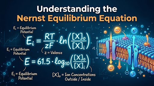
The membrane potential at which net passive flux of that particular ion becomes zero is called the Nernst equilibrium potential for that ion. This equation considers only a single permeable ion in isolation.
The Goldman-Hodgkin-Katz Equation
The Goldman-Hodgkin-Katz equation accounts for multiple diffusible ions (especially sodium, potassium, and chloride) simultaneously to determine the membrane potential.
When an ion moves down its concentration gradient across a selectively permeable cell membrane, it seperates electric charges, creating a transmembrane voltage. But the electrical gradient opposes the concentration gradient with equal force.
The membrane potential at which the net passive movement of that ion is zero is called the Nernst equilibrium potential for that ion. This equation considers only a single permeable ion in isolation.
How to calculate Nernst Equilibrium Equation
The general thermodynamic form of the equation is
Where E= equilibrium potential for the ion; R = universal gas constant
T = absolute temperature; F = Faraday constant; z = valency of the ion.
Converting the natural logarithm to the form 10^-the simple form is
E ion=61.5/z log 10 [ion out]/[log out]
Calculation of Equilibrium Potential for the Potassium,Sodium and Chloride ions
EK=Equilibrium potential for potassium
EK=61.5/ valency of the ion X 40/140= about -94 mV
valency of potassium is one, so it is 61.5 x 40/140 = about -90 m V.
ENa=Equilibrium potential for Sodium
ENa=61.5/ valency of the ion X 142/14= about +61 mV
valency of Sodium is one, so it is 61.5 x 142/14 = about + 61 m V.
ECl=Equilibrium potential for Chloride.
ECl=61.5/ valency of the ion X 105/4= about -86 mV
valency of chloride is one, so it is 61.5 x 10⁵/4= about -86 m V.
Physiological importance:
Resting membrane potential (RMP) of a large myelin nerve fiber is about -70 mV to -94 mV. This is very close to the equilibrium potential of potassium ions.
Cell membranes are highly selective, semipermeable membranes that separate the extracellular fluid compartment (ECF) from the intracellular fluid compartment (ICF).
The compositions of ECF and ICF differ markedly in ionic concentrations. For example, Sodium ions are more abundant in ECF, and potassium ions are more abundant in ICF. Anions like Chloride are diffusible, but many anions are non-diffusible, e.g., proteins, phosphates.
Main points to remember are:
The cell membrane is almost impermeable to Sodium ions.
The cell membrane is almost impermeable to proteins and phosphate ions.
The cell membrane is very permeable to Potassium and chloride ions.
There are many carriers and channels for transport of substances, for example, glucose molecules.
Why are sodium ions with less atomic weight (23) impermeable and potassium ions with atomic weight (39) more permeable?
In the body, ions are present in hydrated form, which alters their effective diameters. In the case of sodium, its atomic weight is 23, but its diameter is 0.18 nm, whereas the atomic weight of potassium is 39, but its diameter is 0.12 nm. Therefore, potassium ions move more easily across the cell membrane.
Gibbs-Donnan Equilibrium and its Clinical importance :
Suppose two cells A and B are separated by a semipermeable membrane. Ions K and Cl are diffusible freely.
Know K in A is equal to K in B: KA = KB and CLA=CLB
And KA = CLA; likewise, KB=CLB;
Product of diffusible ions on A =product of diffusible ions on B
KA × CLA = KB XCLB.
Suppose if cell A has an indiffusable anion P, then
KA= CLA+PA. KA > KB and CLA < CLB.
Therefore, at electrical equilibrium two events occur:
1. The diffusible cation K concentration on the side of the membrane containing the non-diffusible ions P is greater than the diffusible cation K concentration on the other side;
2. The diffusible anion Cl concentration will be greater on the side without the non-diffusible ions.
This is known as Gibbs-Donnan membrane equilibrium.
What are the effects of Gibbs-Donnan :
There will be an electrical difference across the cell membrane. The interior of the cell is negative relative to its exterior. The magnitude of the potential difference in rest depends on its type. We will learn about the Nernst equation.
Because of protein anions and other organic molecules in the cells, there are more osmotically active particles in the cells than the interstitial fluid. This draws water into the cells, causing them to swell and potentially rupture. But sodium-potassium pumps are present in cell membranes, which remove three sodium ions from the cell and allow the entry of two potassium ions into the cell, thereby maintaining osmotic equilibrium between the inside and outside of the cell.
Learning will enrich your knowledge, enabling you to soar high and achieve your maximum potential in life; the sky is the limit.
Caspase8|Caspase9|Smac|Proapoptotic proteins |BAX| BAD BAK| Ceramide | apoptotic bodies| Apoptosis | Necrosis| Cell death|cell programmed death| Regulated cell death
Introduction:
Apoptosis is programmed cell death. It is a well-coordinated and planned cell death procedure. Apoptosis was first described in 1972. Apoptosis is a natural process that is under genetic control.
Apoptosis is different from necrosis.
Apoptosis is derived from the Greek word meaning the falling of leaves from trees or petals. Apo is a prefix meaning “away from,” and ptosis means “falling.”
MechanismofApoptosis:
Apoptosis is triggered by both internal and external stimuli.
Internal stimuli:
Apoptosis-inducing factor is present in the intermembrane space in the mitochondria. Hypoxia, lack of certain hormones, for example, growth hormone, thyroxine, and excess stress radiation cause the release of cytochrome and Smac .’Smac’ is a protein that activates ‘Caspase 9’, which in turn stimulates apoptosis.
Externalstimuli:
y receptors, for example, ‘TNF,’ are present on the cell membrane. Some ligands bind with their specific receptors and provoke a series of reactions leading to activation of ‘Caspase8’. The activated ‘Caspase 8’ causes DNA fragmentation and chromatin condensation.
StepsofApoptosis:
1. Activation of Caspases will cause proteolysis.
2. Hydrolysis of sphingomyelin of the cell membrane forms Ceramide, which causes damage to mitochondria and releases apoptosis-inducing factors.
3. Cell shrinkage
4. DNA damage
5. Liberation of apoptotic bodies and finally
6. Phagocytosis of apoptotic bodies by phagocytic cells of the body.
RegulationofApoptosis:
Apoptosis depends on the delicate balance between:
1. Proapoptotic proteins such as BAX, BAD, BAK, and
2. Antiapoptotic proteins, for example, BCL-2 and Bcl-xL. BCL-2 is the first apoptosis regulatory protein recognized in any organism.
There are many BCL proteins, such as BCL-2, BCL-3, BCL-5, BCL-7, BCL-9, and others.
The synthesis of BCL-2 (B-cell lymphoma-2) is controlled by the BCL-2 gene. BCL-2 is present on the outer membrane of mitochondria and inhibits proapoptotic proteins, promoting cellular survival.
Sometimes it may promote apoptosis by stimulating proapoptotic proteins. Proapoptotic proteins act on the mitochondrial membrane, promoting permeabilization and releasing factors that trigger apoptosis.
Importanceof Apoptosis:
Apoptosis is a normal physiological process that occurs during an organism’s growth and development. This has a vital role in removing harmful and useless cells from the body.
Defects in apoptosis are responsible for malignancy, autoimmune diseases, and organ atrophy.
During fetal development, many tissues degenerate.
Degeneration of the web in the fingers to separate the fingers and toes.
Degeneration of neurons during brain development and formation of synapses.
Thymus involution after childhood. Endometrial shedding in the menstrual cycle. Normal shedding of the intestinal epithelium and some cells. Rejection of transplants.
Damage to the BCL-2 gene is responsible for many cancers, including cancer of the prostate, breast, lungs, melanoma, and some autoimmune diseases. It is also involved in resistance to cancer treatments.
Overactivation of antiapoptotic factors or underactivation of proapoptotic factors is responsible for the reduced cell death that is specific to cancers.
The reverse is also true. Overactivation of proapoptotic factors or underactivation of antiapoptotic factors is responsible for atrophy of organs, neurodegenerative diseases, schizophrenia, Parkinsonism, etc.
Differences between Apoptosis and Necrosis
Necrosis
Apoptosis
1.
Cell size
swells
shrinks
2.
Cell organelle
disintegrate
Remains almost normal
3.
Cytoplasmic changes
homogenously eosinophilic
intensely eosinophilic
4.
Nuclear changes
Condensation and clumping of the nucleus.The disintegration of the nucleus.
Let us learn about Human Physiology and how we humans function
Physiology is the study of the parts of living organisms.
Introduction:
Physiology is the study of the functions of living organisms. It is divided into many branches. We are concerned with Human Physiology. In Physiology, understanding the mechanisms that regulate bodily functions is essential.
Physiology is derived from a Greek word with a Latin equivalent, Physiologia, meaning “natural knowledge”. Physiology is the branch of science that deals with the functions of the body and how these functions occur and are controlled. It also describes which organs initiate and maintain these functions.
A living organism comprises different organs. Body organs function so that mild variations in the external environment do not alter the body’s internal environment, which is kept constant within the normal range.
In Physiology, we study the functions of the living organism. The word Physiology was introduced by Jean Fernel (1497-1558), a French physician. Understanding the mechanisms that regulate bodily functions is essential in physiology.
Physiology is derived from a Greek word meaning “natural knowledge.” The ancient Greek phrase nature origin” and its study indicate an innate scientific understanding of a living body’s station, functions, and mechanisms.
Physiology is the branch of biological science that deals with the body’s functions. It also describes the organs that initiate and control these functions.
Divisions of Physiology:
It comprises many branches.
1. Human Physiology
2. Animal Physiology
3. Plant Physiology
4. Cell Physiology
We are concerned with Human Physiology.
A living organism is composed of many organs that respond to changes in its internal and external environments.
Body organs function so that mild variations in the external environment do not alter the body’s internal environment, which is kept constant within the normal range.
Physiological condition is the condition of normal function, while pathological condition refers to altered physiology-abnormal conditions.
4. Rapid and precise communication between cells occurs in various ways, for example, through chemical and electrical methods.
Animal experiments have provided knowledge of human physiology. Anatomy and physiology are intrinsically related. Physiology is included in medical study globally, and many universities offer bachelor’s, master’s, and Ph.D. degrees in physiology.
Any change in physiology will impact an individual’s physical and mental functions. The study of physiology focuses on the level of organs and systems within the body, and the biological basis of thebody’s operations.
By studying human physiology, we understand how the body maintains normal health and responds to and adapts to challenges in both internal and external environments.
Physiology is a fascinating and dynamic subject that underpins translational and clinical medicine and the interface between the physical and life sciences.
Developments:
Documents reveal that the study of medical or human physiology began in Greece in the late 5th century BC, with Hippocrates. Around the same time, it also started in India and some other countries.
Later, many scientists, philosophers, and thinkers contributed to the development of physiology. Some of them are Galen, Aristotle, Claude Bernard, Walter B. Cannon, and many others.
Scopes in physiology
Anatomy and physiology are intrinsically related. Physiology is included in medical study globally, and many universities offer bachelor’s, master’s, and Ph.D. degrees in physiology.
Physiology is an essential subject in medical education for M.B.B.S. degrees. It is crucial in other systems, such as B.Sc. M.Sc. physiology.
A physiologist may be posted in medical colleges as a tutor, assistant professor, associate Professor, or Professor.
Exercise physiology is the study of physical activity. The word exercise comes from the Latin word drive forth’’
Exercise has been considered important to human health for thousands of years in ancient cultures. The Greek physician Hippocrates supported exercise and recommended moderate activity to maintain health and improve it.
Many careers in exercise physiology are available, and job opportunities are expected to increase as the population ages and obesity rates continue to rise. An exercise physiologist may pursue research work.
Physiologists can work in various clinical or non-clinical settings,
including fitness centers, community organizations, and corporate fitness.
Sports physiologists may work in private fitness clinics. They may start their own clinic.
Clinical physiologists may be employed by hospitals, community facilities, industries, and nursing homes.
The thymus is a small (5 mm x 5mm) gland, a central or primary lymphoid organ. It has two lobes that are connected by a fibrous isthmus.
Location;
It is situated in the superior mediastinum and extends upward to the root of the neck and downward to the anterior mediastinum.
Location of the thymus
Structure of the thymus
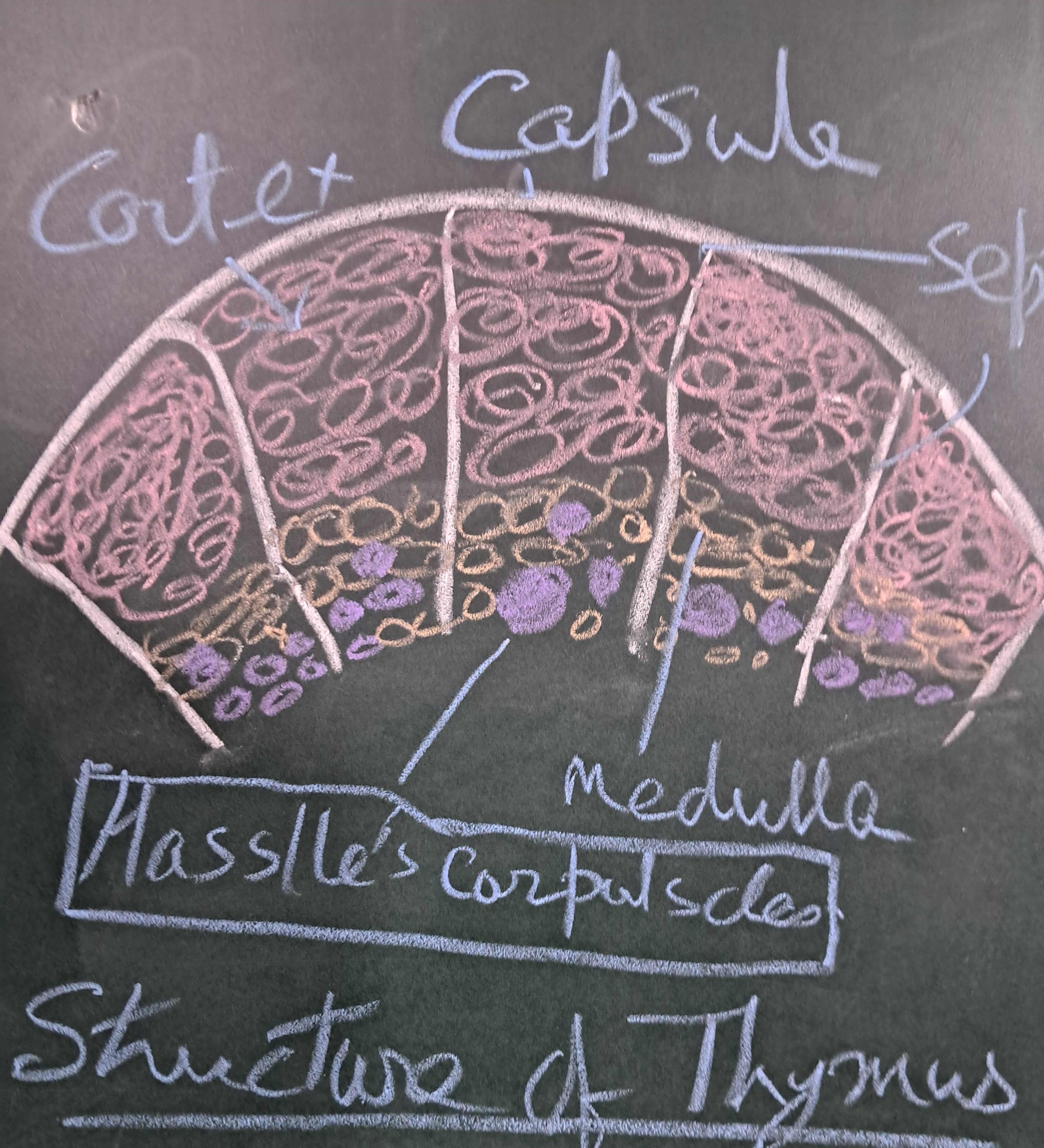
A capsule covers the thymus and sends septa into the organ and divides it into lobules. Each lobule consists of an outer cortex and an inner medulla.
Cortex:
Structure of the Thymus
1. A densely populated area where actively multiplying lymphocytes are present, with immature T-lymphocytes or thymocytes. The cells undergo differentiation.
2. Epithelial reticular cells (TECs- Types I, II, III) are present that give structural support.
3. Macrophages phagocytose apoptotic T-cells.
Medulla
It is pale in color and contains
1. A few more mature T-lymphocytes
2. Specialized epithelial cells (Types IV, V, VI)
3. Hassall’s corpuscles or thymic corpuscles are characteristic of the thymus. A central calcified core is surrounded by concentrically arranged keratinized epithelial reticular cells.
Secretion and Functions
Epithelial reticular cells produce endocrine and paracrine effects vital for T-cell differentiation.
Thymosin and Thymulin promote T-cell proliferation and differentiation.
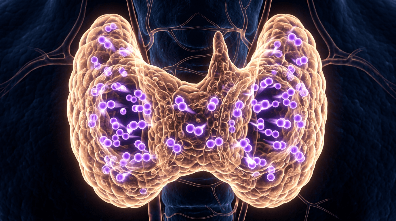
The thyroid gland is a butterfly-shaped endocrine gland located superficially in the neck. It consists of two lobes, the right and left lobes, connected in the lower two-thirds by a thin band of tissue known as the isthmus.
Shape: It is a butterfly-like endocrine organ. In the 1600s, Thomas Wharton named this gland for its resemblance to an ancient Greek shield, or ‘thyos.’
Size: The gland weighs 25 grams. Each lobe is 5 cm long, 3 cm wide, and 2 cm thick. The isthmus is 1.25 cm in height and breadth. The thyroid gland is more prominent in women than in men and increases during pregnancy or any other condition with anxiety and tension. It is very vascular and the largest endocrine gland.
Location: The thyroid gland is present in front of the larynx and trachea.
Relation of the Thyroid Gland:
Above: The thyroid and cricoid cartilages lie above it.
Below: The Adam’s apple lies below the gland.
Front: The infra-thyroid muscles lie in front.
Behind: The larynx, lower pharynx, and esophagus lie behind it.
On the sides: The sternocleidomastoid muscles and carotid arteries are present bilaterally.
Four parathyroid glands are present, two on each side. They lie between the two layers of the thyroid capsule at the back of the thyroid lobes.
A thin, two-layered fibrous capsule covers the gland. The inner layer invaginates the glandular mass, forming septa that divide the thyroid tissue into macroscopic lobes. The outer layer fuses inferiorly and continues into the pretracheal fascia, forming the posterior suspensory ligament of the thyroid gland. This ligament is known as Berry’s ligament.
Due to this ligament, the thyroid gland moves with the cartilages during swallowing.
Blood supply
Ø Arterial blood supply is by the superior and inferior thyroid arteries.
Ø Venous blood drainage: Through the superior, middle, and inferior thyroid veins.
Lymphatic drainage
The lymph drains into the pre-laryngeal, pre-tracheal, and paratracheal lymph nodes.
Nerve supply:
Ø Sympathetic nerve supply: From the superior, middle, and inferior cervical ganglia of the sympathetic trunk.
Ø Parasympathetic nerve supply: via the superior and recurrent laryngeal nerves.
Variations
There are many variations. For example, a third lobe is sometimes present, known as the ‘pyramidal lobe’ or ‘Lalouette’s pyramid.’
Development of the gland
At three weeks of gestation, the gland develops in the pharyngeal floor at the base of the tongue. It then grows in front of the pharynx and migrates to the bottom of the neck over the next few weeks. During migration, it remains connected to the tongue by a narrow tube known as the thyroglossal duct. At the end of the 5th week, the duct degenerates, and over the following two weeks, the detached gland migrates to its final position. In some cases, the thyroglossal duct remains patent even in adulthood.
Histology of thyroid gland:
The thyroid is made up of multiple small follicles. A follicle measures only 0.02-0.9 mm. About 40 small follicles form a lobule. A single layer of cuboidal cells surrounds the lumen of a follicle. When the gland is active, cuboidal cells change into columnar cells; when the gland is inactive, they become flat epithelial cells. The follicular cell is also known as the thyrocyte.
Some neuroendocrine parafollicular cells, also known as C cells, are present among the follicular cells and secrete calcitonin.
Secretion of the thyroid gland
The thyroid gland secretes three hormones:
(i) Thyroxine (T4)
(ii) Tri-iodothyronine (T3), and
(iii) Calcitonin.
The follicular lumen is filled with a transparent, gelatinous, pink colloid containing iodine, known as thyroglobulin.
Thyroid cells are highly active and contain abundant mitochondria, rough endoplasmic reticulum, and a Golgi apparatus. Their microvilli project into the follicular lumen. They synthesize protein and secrete it into the lumen by exocytosis. The rough endoplasmic reticulum of thyrocytes synthesizes thyroglobulin, which enters the follicular lumen by exocytosis. It is stored as a colloid and reacts with iodine in the follicular lumen.
Iodine metabolism
Iodine is a trace element essential for thyroid hormone production. In blood, iodide (I-) is present. Thyrocytes absorb iodides against the electrical gradient through an ion channel. In the sodium-iodide symporter channel on the cell membrane, two sodium ions move out of the cell, and one iodide ion moves into the cell. Finally, the iodide passes from the cell into the follicular lumen through pendrin, an iodide-chloride antiport.
In the follicular lumen, iodide is oxidized to iodine and becomes very active. In the presence of thyroid peroxidase, tyrosine is activated, and the enzyme combines with it to form MIT and DIT.
At first, MIT (monoiodotyrosine) is formed, then DIT (diiodotyrosine).
MIT+MIT=DIT
DIT plus MIT equals Triiodotyrosine (T3).
DIT+DIT=Tetraiodotyrosine (T4).
The follicular cells reabsorb thyroglobulin from the lumen. The iodinated tyrosines are cleaved, yielding MIT, DIT, T3, T4, and reverse triiodothyronine.
T3 and T4 enter the blood. In blood, 80-90% is T4 and 20-30% is T3.
MIT contains one atom of iodine per molecule, DIT contains two iodine atoms per molecule, T3 contains three iodine atoms per molecule, and T4 contains four iodine atoms per molecule.
Only a small percentage of thyroid hormone (0.3%) is free and active. In circulation, T4 produces 85% of T3.
70% are bound to thyroxine-binding globulin.
15% are bound to albumin, and
10% are bound to transthyretin.
C cells present between the follicular cells secrete calcitonin, which regulates blood calcium concentration. When blood calcium levels rise, calcitonin decreases calcium release from bone and increases its absorption. It helps regulate blood calcium levels.
1. Reduces the release of calcium from bone.
2. Increases calcium absorption by bone.
Calcitonin is not as potent as the parathyroid hormone secreted by the gland and opposes the effect of parathyroid hormone.
Mechanism of action
Thyroid hormones enter cells by crossing cell membranes and binding to nuclear thyroid hormone receptors TR-α1 and α2, and TR-β1 and β2.
These receptors bind with hormone response elements and transcription factors to modulate DNA transcription.
In addition, thyroid hormones act with various enzymes in the cytoplasm, including calcium ATPase, adenylyl cyclase, and glucose transporters.
Functions of the thyroid gland
Hormones of the thyroid gland are not essential for life, but their removal in adults leads to impaired temperature regulation and mental and physical health problems.
The thyroid hormones have many functions and affect all body tissues. These include
Thyroid hormones are especially required for normal fetal CNS development and fetal bone maturation.
The thyroid hormones
Ø Increase the basal metabolic rate and protein synthesis.
Ø Stimulate oxygen consumption of most body tissues, help regulate fat and carbohydrate metabolism, and are essential for tissue differentiation.
Ø Stimulate absorption from the gut, uptake by cells, breakdown of glucose, breakdown of fats, increase the number of free fatty acids and thyroid hormones, and decrease cholesterol levels.
Ø The cardiovascular effect is to increase the rate and strength of the heartbeat, increasing the rate of breathing, intake, and consumption of oxygen.
Ø The thyroid hormones are essential for maintaining normal sexual functions, including libido and a regular menstrual cycle.
Ø During development, a deficiency of thyroid hormones in children will cause intellectual disability.
Euthyroid indicates normal thyroid function.
Hyperthyroidism: when there is an excess of thyroid hormones.
Hypothyroidism: when there is a low amount of thyroid hormone.
Thyroiditis: when there is inflammation of the thyroid gland.
Goiter: when there is an enlargement of the thyroid gland.
Other conditions related to the thyroid glands are:
Thyroid nodules, thyroid cancer, autoimmune disorder Graves’ disease, etc.
The gland and its various diseases have been noted and treated for centuries.
How are these hormones metabolized?
Deiodinase enzymes in peripheral tissue remove iodine from MIT and DIT, converting T4 to T3 and RT3.
Thyroid hormones exert negative feedback on TSH and CRH. High levels of thyroid hormones suppress TSH production.
The study of the relationship between bladder volume and intravesical pressure is called cystometry, which measures the bladder’s contractile force during urination.
Cystometry can help diagnose problems with bladder filling and emptying. Also known as flow cystometry, it evaluates bladder function — the capacity to contract and expel urine.
Cystometrogram
A cystometrogram (CMG) is a plot of volumes against intravesical pressures. In other words, a Cystometrogram plots the volume of urine passed from the bladder against intravesical pressure.
Importance
Cystography can diagnose bladder problems.
By cystoscopy, we can inspect the interior of the bladder.
The spleen is the largest wedge-shaped, dark purple-colored lymphatic organ of the lymphoid system, and it is connected to the vascular system. It is highly vascular. Its structure is similar to a large lymph node.
Measurements –length-5inches, breadth-3 3 inches,thickness-1 inch. Weight(average) is only 7 ounces.
Spleen performs multiple functions, although it is not essential for survival ,it is vital for healthy life. Dysfunctions of spleen will produce serious problems.
Location
The left hypochondrium houses the spleen between the fundus of the stomach and the diaphragm.
Behind the mid-axillary line opposite the 9th,10th, and 11th ribs.
External features Macroscpic Feature
Two ends, two surfaces, two angles, and three boundaries.
Two ends: (1) anterior or lateral end that faces downward and forward, (2) posterior or medial face that faces upward and backward.
Three boundaries: 1. superior,2.inferior, and 3. intermediate.
The superior border is convex and faces and is in contact with the undersurface of the left side of the diaphragm. This has one or more notches called the splenic notch.
The inferior border is rounded and corresponds to the lower border of the eleventh rib.
The intermediate border is round and separates the gastric impression from the renal impression.
Two surfaces-1. The diaphragmatic surface is smooth, convex, and directed upward, backward, and to the left.
1. Visceral surface is concave and irregular and has four impressions 1. Gastric impression is the most significant impression and is due to the fundus of the stomach. It lies between the superior and intermediate border of the spleen.
Renal impression is due to the left kidney. It is present below and behind the gastric impression and lies between the intermediate and inferior boundaries.
The left colic flexure produces a colic impression. It is triangular and situated in front of the lateral end. and
The tail of the pancreas causes the pancreatic impression, which is located between the hilum and the colic impression.
There is a groove through which the splenic artery and nerves enter, and veins and lymphatic exit.
Relation of the Spleen
The spleen is surrounded by peritoneum and suspended by four ligaments.
1. Gastrosplenic ligament- This ligament extends from the hilum of the spleen to the upper one-third of the greater curvature of the stomach. Short gastric vessels are present in this ligament.
2. Splenorenal ligament- This ligament extends from the hilum of the spleen to the anterior surface of the left kidney. Tail of the pancreas, splenic venules, and pancreatosplenic lymph nodes are present in this ligament.
3. Colicosplenic ligament
4. Phrenocolic ligament
The visceral surface of the spleen is related to the fundus of the stomach, the anterior surface of the left kidney, the left colic flexure, and the tail of the pancreas.
Near the middle of the spleen, a long fissure is present, which is known as the hilum.
Penicilliary radicles are arterioles that supply germinal centers.
Develops from mesenchymal tissue, while most of the gut organs are derived from endoderm.
Histology
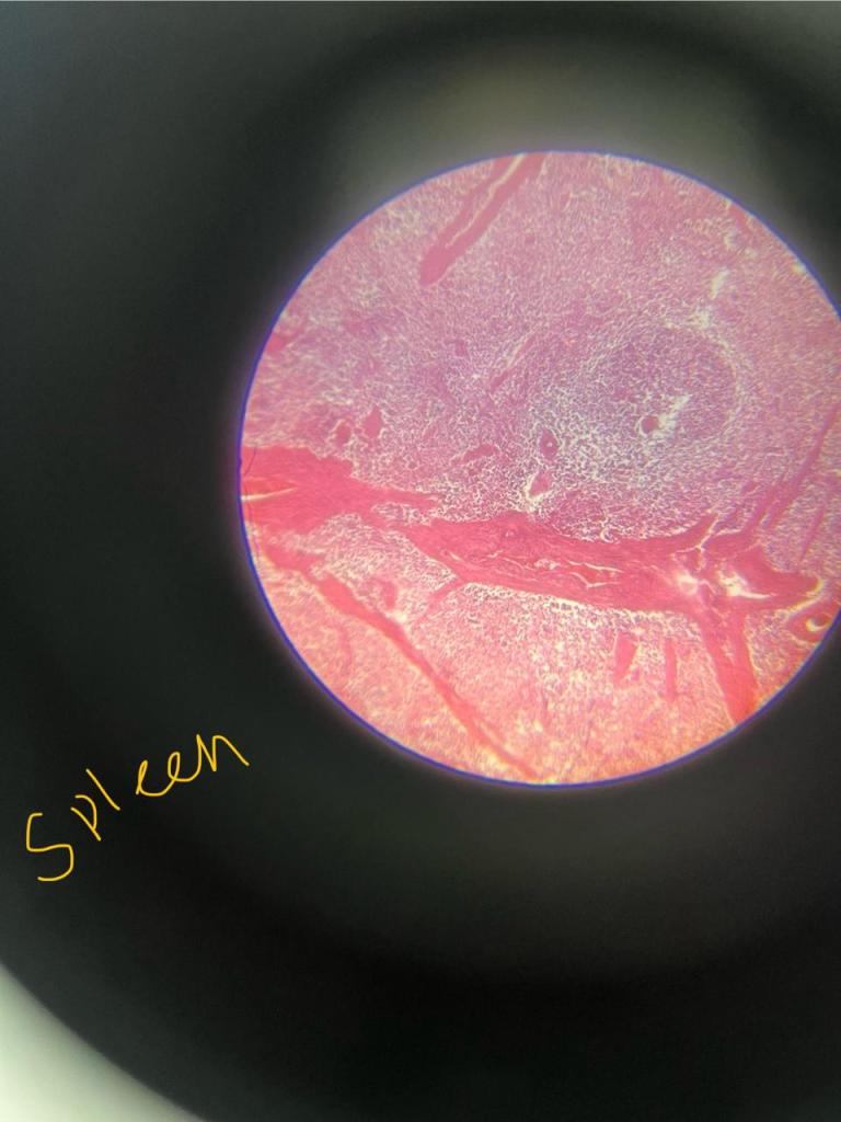
The outer capsule covers the spleen, made up of thick fibrous tissue.
Beneath the capsule, there is a subcapsular space. The spleen contains two different types of tissues, white pulp and red pulp.
The white pulp is white in colour and contains growing immune and blood cells. White pulp is composed of Malpighian corpuscles. These include lymphoid follicles rich in B lymphocytes and periarteriolar lymphoid sheaths rich in T lymphocytes.
The red pulp is red in colour and contains sinuses filled with blood, reticular fibers, and a marginal zone bordering on the white pulp.
Blood supply
The Splenic artery, the largest branch of the coeliac artery, provides the main blood supply. At the hilum, it divides into five or more branches and enters the spleen. Within the spleen, it divides repeatedly into straight arterioles called Penicilliary radicles. The penicilliary radicles supply the germinal centers.
Blood supply of the Spleen
Close theory: capillaries open directly in the venous sinusoids and lie in the red pulp.
Open theory: capillaries open into red pulp from where blood enters sinusoids through their fenestrated wall.
Compromise theory: according to this theory, circulation is open in the distended spleen and closed in the contracted spleen.
Lymphatic drainage
The Spleen has only efferent lymphatic vessels. There are no lymphatics in the proper splenic tissue. The tissue fluid formed in the spleen freely enters the venous sinusoids. However, the splenic lymphatics are present in the capsule, trabeculae, and visceral peritoneum, and drain into the pancreatocoliecal lymph nodes.
Venous drainage
The splenic vein is formed by the confluence of venules at the hilum, which join the superior mesenteric vein to form the portal vein. Its tributaries are 1. the short gastric vein, 2. the left gastroepiploic vein, 3. the pancreatic vein, and inferior mesenteric vein.
Nerve Supply
Sympathetic fibre derived from the celiac plexus supplies smooth muscles present in the blood vessels, as well as in the trabecular cord.[ splenic plexus, which connects a branch of the celiac ganglion to the vagus nerve.
Functions
1. Filtration of blood
2. Reservoir of blood cells
3. Active immune response: cell-mediated and humoral immunity.
4. Produces opsonins, properdin, and tuftsin.
5. Hematopoiesis in the embryo and in case of emergency.
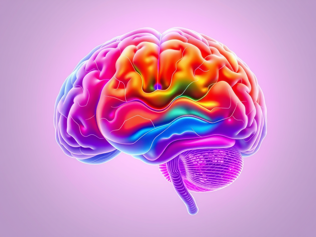
The limbic system is derived from the word ‘Limbus’, meaning a ring. The word limbic was first used to describe the border structures around the basal region of the cerebrum.
Now the limbic system describes the entire neuronal circuitry that controls emotional, behavioral, and motivational drives and more.
Components of the Limbic System
1. The limbic lobe and the related subcortical nuclei.
The limbic lobe includes the cingulate gyrus, isthmus, hippocampal gyrus, and uncus.
2. The related subcortical nuclei are the hypothalamus, amygdala, septal nuclei, portion of the basal ganglion,paraolfactory area, and anteriorthalamic nucleus.
The hypothalamus is located in the middle of all these structures and plays a vital role in the limbic system.
The limbic cortex surrounds these subcortical limbic areas. The limbic cortex is composed of a ring of cerebral cortex on each side of the brain. The limbic cortex begins.
1. In the orbitofrontal area on the ventral surface of the frontal lobes,
2. Extends upwards into the subcallosal gyrus,
3. Then over the top of the corpus callosum onto the medial aspect of the cerebral hemisphere in the cingulate gyrus, and finally
4. Passing behind the corpus callosum and downward onto the ventromedial surface of the temporal lobe to the parahippocamal gyrus and uncus.
The neocortex sits with the legs on each side of the limbic system, like a rider on a horse without complete control. It can modify emotional behavior, but cannot initiate or terminate emotional behaviors.
Histology
The limbic cortex is made up of Allocortex (allo means old). The limbic cortex is the phylogenetically oldest part of the cerebral cortex.
Connections
The limbic cortex functions as a two-way communication and association linkage between the neocortex and the lower limbic structures.
1. Reticular nuclei in the brainstem and their associated nuclei
2. Medial forebrain bundle- main
3. Through short pathways among the reticular formation of the brainstem, thalamus, hypothalamus, and other areas of the basal brain.
Functions
1. Controls autonomic functions
2. Emotional behaviours like pleasant or unpleasant, reward or punishment, satisfaction or aversion.Emotion of rage, fear.





Leave a Reply