
Table of Contents
Introduction:
The thymus is a small (5 mm x 5mm) gland, a central or primary lymphoid organ. It has two lobes that are connected by a fibrous isthmus.
Location;
It is situated in the superior mediastinum and extends upward to the root of the neck and downward to the anterior mediastinum.

Location of the thymus
Structure of the thymus
A capsule covers the thymus and sends septa into the organ and divides it into lobules. Each lobule consists of an outer cortex and an inner medulla.
Cortex:
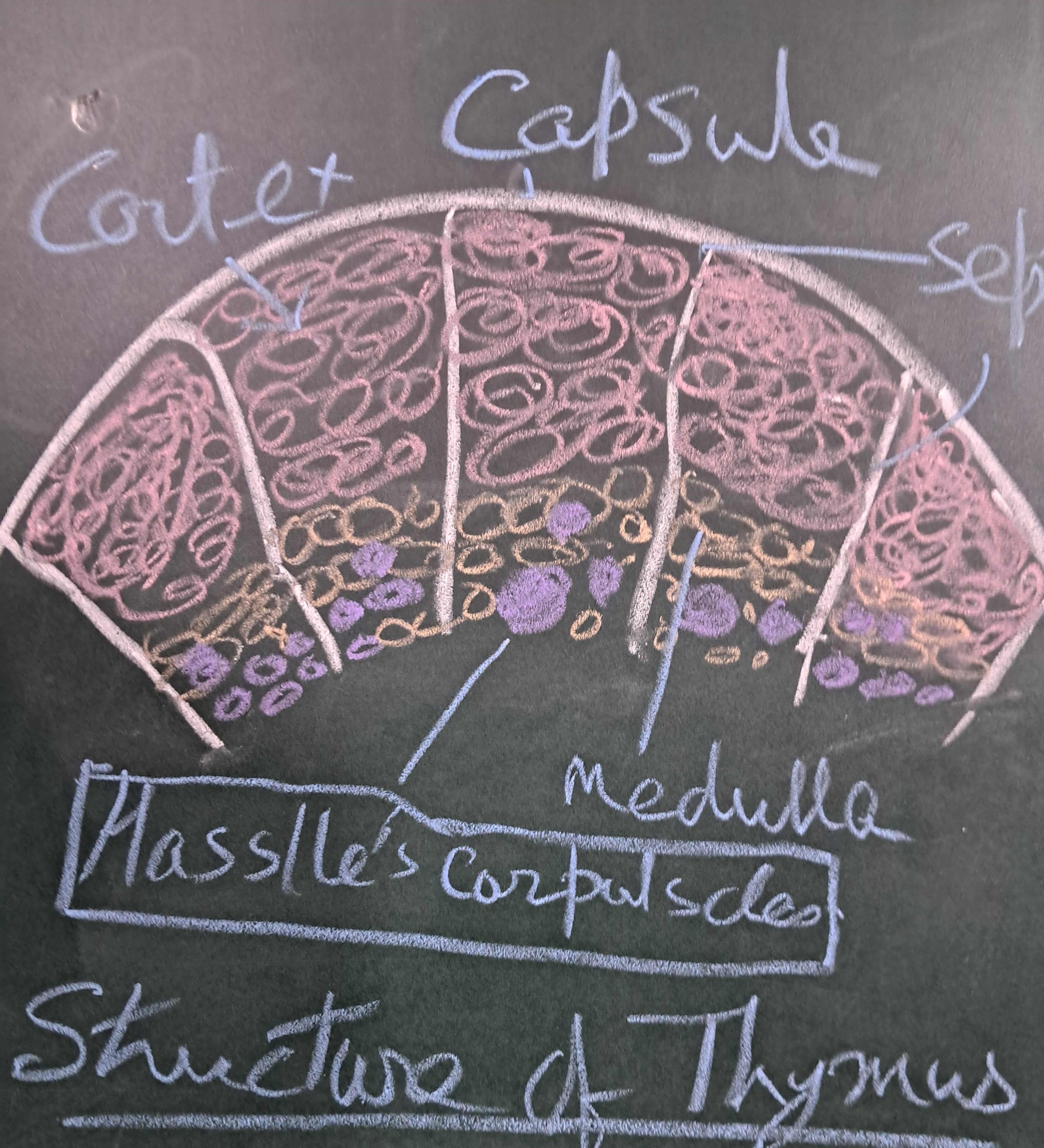
1. A densely populated area where actively multiplying lymphocytes are present, with immature T-lymphocytes or thymocytes. The cells undergo differentiation.
2. Epithelial reticular cells (TECs- Types I, II, III) are present that give structural support.
3. Macrophages phagocytose apoptotic T-cells.
Medulla
It is pale in color and contains
1. A few more mature T-lymphocytes
2. Specialized epithelial cells (Types IV, V, VI)
3. Hassall’s corpuscles or thymic corpuscles are characteristic of the thymus. A central calcified core is surrounded by concentrically arranged keratinized epithelial reticular cells.
Secretion and Functions
Epithelial reticular cells produce endocrine and paracrine effects vital for T-cell differentiation.
Thymosin and Thymulin promote T-cell proliferation and differentiation.
Thymopoietin regulates T-cell differentiation.
Thymic Humoral Factor enhances cell-mediated immunity.
Hassall’s corpuscles secrete TLSP to induce regulatory T-cells for central tolerance.
Clinical
Thymoma or thymic carcinoma usually causes pressure syndrome.
Ectopic thymic tissue.
Associated with Myasthenia Gravis and DiGeorge syndrome.


Leave a comment