Hemopoiesis

Hemopoiesis

The formation of formed elements of blood is hemopoiesis. There are three types of formed elements of blood-Erythrocytes[RBC], leucocytes[WBC], and Thrombocytes [platelets].

  1. Hemopoiesis
    1. Types of hemopoiesis
  2. Site of erythropoiesis
    1. The mesoblastic stage
    2. The hepatic stage
    3. The myeloid stages
  3. Stages of erythropoeisis are:-
  4. Main features In erythropoiesis –

Types of hemopoiesis

So, hemopoiesis is also of three types –

Erythropoiesis-formation of erythrocytes [RBC].

Leucopoiesis -formation of leucocytes [WBC],

Megakaryocytopoiesis – formation of thrombocytes [platelets].

Site of erythropoiesis

Erythropoiesis is the process of erythrocyte formation; it is a continuous process. However, at different ages, the site of erythropoiesis differs. The sites :

It may be extravascular– hepatic and myeloid stages.

It may be intravascular – mesoblastic stage.

The mesoblastic stage

The mesoblastic stage of erythropoiesisstartsin theearly embryo and lasts up to three months of fetal life. RBC is formed from the ‘area vasculosa’-mesoderm of ‘yolk sac.’ The mesoderm of the yolk sac consists of a nucleated mass of protoplasm that creates a network of capillary vessels lined by endothelium and filled with plasma. Endothelial cells proliferate and form masses of nucleated cells. These nucleated cells contain hemoglobin. And detach from capillaries. Later, these cells lose their nuclei and become non-nucleated red blood cells.

The hepatic stage

The hepatic stage of erythropoiesisstarts afterthree months of fetal life and lasts until six months of fetal life in the liver and spleen. Then, RBCs are formed from the mesenchyme between the tissue cells and the blood vessels.

The myeloid stages

Myeloid stages of erythropoiesis start in the bone marrow after six months of fetal life. If the bone marrow fails to form RBCs, the liver and spleen will start erythropoiesis.

At a young age, erythropoiesis occurs in red marrow and is present in almost all bones.

In old age, erythropoiesis occurs only in the red marrow of flat bones like the sternum, iliac crest, and vertebrae.

In myeloid erythropoiesis, red blood cell formation occurs from committed stem cells and takes about seven days to mature.

Pluripotent stem cells of the bone marrow develop into Lymphoid stem cells and myeloid stem cells.

Now myeloid stem cells will form ‘colony forming unit EM’ [Erythrocyte and megakaryocyte] and ‘colony forming unit GM ‘[Granulocyte myelocyte].

The ‘colony-forming unit EM’ will form colony-forming units CFU-E’ and CFU-M.

CFU E will form proerthroblast, which [blast is rapidly growing

cells], proliferating to form erythroblast. A proerthroblast is a large cell of 15 -20 microns with a large nucleus and 3-4 nucleoli. Chromatin is open-eukaryotic. It is a highly basophilic and rapidly dividing cell.

The size of the erythroblast is less than the proerythroblast [15-18 microns] and is less basophilic cell. The nucleus also decreases in size and loses nucleoli. Chromatin condenses. It also divides. This erythroblast is known as basophilic erythroblast or early normoblast.

The polychromatic erythroblast is 10-15 microns in size. The nucleus decreases in size, and chromatin condenses. Hemoglobin appears in the cytoplasm, giving it a red color. The ribosome is still present in the cytoplasm, showing a blue color. So, this cell is known as a polychromatic erythroblast and intermediate normoblast.

The size of the orthochromatic erythroblast is 8 to 10 microns. This is also known as the late normoblast. The orthochromatic erythroblast is red due to the cytoplasm’s large amount of hemoglobin. Some amount of ribosomes is also present. The nucleus becomes very small -looks like a cartwheel.

[These stages of erythrocyte development occur in the bone marrow and take about seven days in normal conditions.]

A reticulocyte is a small cell [7 to 8 microns]without a nucleus. Some RNA presently gives a reticular appearance to the cell. Only 1-2% of reticulocytes are present in the circulation. However, in rapid erythropoiesis, reticulocyte percentage increases in circulation. Reticulocyte is also known as ‘immature red blood cell.’

.After about two days, reticulocytes become erythrocyte-mature red blood cells.

Erythrocyte is also a tiny cell [7-8 microns] without a nucleus and RNA remnants. It is red.

Hemoglobin appears in the intermediate normoblast stage.

Mitosis stops in the late normoblast stage.

The nucleus and all organelles, such as mitochondria, Golgi apparatus, ribosomes, and endoplasmic reticulum, disappear in the later part of the late normoblast stage.

Reticulocyte maturation continues even after it loses its nucleus and other organelles. The reticulocyte loses 20 to 30 % of the cell surface and eliminates membrane-bound cytosolic organelles through an autophagy /exosome-combined pathway.

Stages of erythropoeisis are:-

Stages Size Other feature

1. Proerythroblast 15-20 µm large nucleus

2. Early normoblast 14-18 µm hemoglobin appears

3. Intermediate normoblast– 10-14 µm more hemoglobin appears

4. Late normoblast – 8-10 µm nucleus disappears

5. Reticulocyte – 7.2 µm reticular structure in the cytoplasm

6.Erythrocyte – 7.2 µm

Stages 1 to 5 are present in the bone marrow. One to two percent of reticulocytes are currently in circulation. In rapid erythropoiesis, the number of reticulocytes increases in circulation. Stage 6, that is, erythrocytes are present only in circulation.

Main features In erythropoiesis –

The size of the cell reduces progressively.

The nucleus decreases in size, and nucleoli disappear as maturation occurs. Open chromatin in the nucleus condenses and finally disintegrates. The nucleus and nucleoli degenerate.

Hemoglobin appears in the intermediate normoblast stage and increases progressively.

Mitosis takes place up to the intermediate normoblast.

Image created by Author with Canva.

Oxytocin |Love Hormone |Cuddle Hormone

Introduction

The lecture explains the site of secretion, functions, and mode of action of oxytocin. It will also explore the ‘milk let down reflex’ or suckling reflex.

Keywords: Oxytocin |Love Hormone | Neurophysin -1|Paraventricular nucleus|peptidergic neurons of the magnocellular neurosecretory system |Milk ejection reflex |Milk let down reflex|Oxytocin and sex|Oxytocin and breastfeeding|Oxytocin benefits for men| Oxytocin benefits|benifits o Oxytoin| Action of oxytocin

Table of contents

  1. Introduction
  2. Table of contents
  3. What is Oxytocin?
  4. Action of oxytocin
  5. Control of oxytocin secretion:
  6. Milk ejection reflex or Milk let-down reflex
  7. Mechanism of action:
  8. Your queries

About’ totalphysiology.com.’

This article is part of my mission to provide trustworthy recent health information to support the general public, patients, and professionals globally.

Here, you will find human Physiology and health-related topics.

This activity aims for learners to better apply the latest scientific knowledge.

Upon completing the article, you will have increased knowledge regarding the subject and use it with great confidence.

What is Oxytocin?

Oxytocin is a hormone and a neurotransmitter.

Oxytocin is secreted mainly from the paraventricular nucleus of the hypothalamus. It is also synthesized in peptidergic neurons of the magnocellular neurosecretory system or the hypothalmo-hypophysial neural tract.

From the paraventricular nucleus, the hormone passes to the axons, combines with the carrier protein ‘Neurophysin -I,’ is transported in the hypothalamus-hypophysial neural tract, and is stored in axon endings in the posterior pituitary.

In the posterior pituitary, oxytocin is stored and released by nerve impulses in the hypothalmo-hypophysial neural tract.

The Love hormone

Oxytocin, also known as the Love hormone, is responsible for positive emotions, social binding, love, relationships, pair bonding, and trust.

Oxytocin causes attachments between mothers and their infants and children and bonds between romantic partners. 

Do you know what is the Cuddle hormone?

Let me know in the comment section.

What does oxytocin do?

Action of oxytocin

1. On the myoepithelium lining the ducts of mammary glands:

It causes contraction of the myoepithelium lining the mammary glands’ alveoli, ducts, and cisternae, leading to milk ejection from the lactating breast.

2. On the myometrium

It stimulates the contraction of the uterus’s smooth muscles, making it vital and helpful in inducing labor. It helps to control bleeding after delivery(PPH). Pitocin is the brand name of Oxytocin injection. It is used to control bleeding after delivery(PPH).

3. Action on the anterior pituitary gland

Oxytocin stimulates the release of lactogenic and galactopoietic factors from the anterior pituitary gland.

4. On blood vessels

In high doses, it causes relaxation of blood vessels, causing a fall in blood pressure.

6. Boosts the prostaglandin production.

5. In males, it increases contraction of the vas deferens’ smooth muscles, facilitating the transport of sperms towards the urethra. The testis, epididymis, and prostate glands in males have oxytocin receptors.

6. Mental health: Oxytocin promotes positive feelings, increases happiness, and promotes positive social behavior. It also plays a role in anger management. A low oxytocin level is associated with depression.

Control of oxytocin secretion:

1. Factors increasing oxytocin secretion:

It may be conditioned sight; crying of the child can cause milk ejection.

Physical stimulation of the nipple.

Physical stimulation of the genital tract.

Dilatation of the cervix also stimulates the paraventricular nucleus to secrete and release oxytocin.

Stimulation of cholinergic nerve.

How to increase oxytocin levels?

You can increase your Oxytocin levels with simple yet very effective steps, such as eating a healthy, balanced diet, exercising regularly, giving someone a hug, and spending time with friends, children, and family.

Touch and massage also increase oxytocin levels.

No food supplement is required to increase oxytocin levels.

2. Factors decreasing oxytocin secretion:

Drugs

Sympathetic nerve stimulation

Stress and psychic factors.

Milk ejection reflex or Milk let-down reflex

Tactile receptors are present in the alveolar region of the breast. They are stimulated by suckling and transmit signals to the paraventricular nucleus, which causes reflex secretion of oxytocin in the blood.

Oxytocin acts on its specific receptors present on the myoepithelium lining the alveoli, ducts, and cisternae of the mammary glands and causes contraction of the myoepithelial lining. This leads to milk ejection from the lactating breast in 30 to 60 seconds.

Suckling also promotes prolactin stimulation by inhibiting the release of ‘prolactin inhibitory factor from the hypothalamus.

In this way, suckling causes both secretion and ejection of milk.

Mechanism of action:

When oxytocin binds to its specific receptor, it activates an enzyme, G2, which stimulates Phospholipase C, which is present on the inner surface of the cell membrane.

Phospholipase C catalyzes the hydrolysis of (PIP2) Phosphatidylinositol diphosphate to yield two second messengers: (IP3) Isositol triphosphate and (DAG) diacylglycerol.

Inositol triphosphate (IP3) diffuses to the endoplasmic reticulum to trigger the release of Ca++ into the cell. The calcium influx increases intracellular calcium ions, leading to muscle contraction.

This mechanism operates in the mammary gland, uterus, and vas deference.

Diacylglycerol stays in the cell membrane to activate protein kinase C.

Your queries:

What is Oxytocin?

What does oxytocin do?

How are oxytocin levels controlled?

How to increase oxytocin levels?

How can oxytocin affect mental health?

Is oxytocin a happy hormone?

Why is oxytocin called the love hormone?

Brand name of oxytocin?

What is the Cuddle hormone?

Oxytocin is the Cuddle hormone. It is associated with pair bonding, sexual activity, childbirth, and breastfeeding. It is also responsible for sexual desire, erection, orgasm, and happiness.

Internal Links:

https://blog.totalphysiology.com/2024/10/growth-hormone-somatotrophin-human.html

https://blog.totalphysiology.com/2022/02/hormones-adrenal-medulla-adrenalin.html

https://blog.totalphysiology.com/2022/01/pituitary-gland secretion hormones.html

External Links: 

Please submit your comments about this article. The team will work hard to evaluate the statement and make appropriate corrections. Your comments will help improve the content.

Thank you very much for reading. I appreciate that you spent some time with us. If you enjoyed reading, please leave a review or a comment. Your comments will encourage other folks to read.

Disclaimer: All possible measures have been taken to ensure the accuracy and reliability of the information; however, the author does not take any liability for using any information provided by the website solely to the viewers. ‘The information is provided as an educational service and public awareness. It is not medical advice. We advise you to review a reference book in case of any doubt and more accurate and advanced knowledge

  1. Introduction
  2. Your queries:

What Happens to the Fat After Its Absorption?

After the fat is absorbed, the chylomicrons enter the liver and tissues, where they are utilized. Fat circulates in the blood and enters the liver and cells.

Table of contents

  1. Fate of fat in circulation
  2. Fate of fat in the tissues
  3. Fate of fat in the liver
    1. Phase I metabolism of fat
    2. Phase II metabolism of fat.
    3. Phase III metabolism of fat.
  4. Role of the liver during carbohydrate deficiency
  5. β-oxidation of fatty acids.

Fate of fat in circulation

Lipoprotein lipase acts on the circulating chylomicrons, VLDL, and triglycerides to make free fatty acids and glycerol. This lipolysis reaction remarkably increases adipose cells’ free fatty acid and glycerol content.

Feeding increases Lipoprotein lipase activity. Therefore, more free fatty acids and glycerol enter the fat cells after feeding.

Fasting and stress decrease Lipoprotein lipase activity, so little free fatty acids and glycerol are available for the fat cells.

Fate of fat in the tissues

Hormone-sensitive lipase catalyzes stored triglycerides into free fatty acids and glycerol. As its name suggests, it is hormone-dependent.

Growth hormone, glucagon, cortisol, epinephrine, and norepinephrine increase activity.

Insulin, prostaglandin E, and feeding decrease its activity.

In this way, we observe that feeding favors fat accumulation in the adipose cells.

Fate of fat in the liver

As chylomicrons enter the liver, ‘liver lipase’ breaks it into free fatty acids and glycerol. This is a very quick reaction.

Phase I metabolism of fat

Triglycerides————–free fatty acids + glycerol.

The glycerol is utilized in the carbohydrate pathway. Glycerol in the carbohydrate metabolism may combine to form fats.

Phase II metabolism of fat.

The fatty acids are oxidized to ‘acetyl-co A’ by β-oxidation of fatty acids.

Phase III metabolism of fat.

‘Acetyl-CoA’ is usually wholly oxidized to form CO2, H2O, and energy.

‘Acetyl-CoA’ may form pyruvic acid and may form carbohydrates.

Excess ‘Acetyl-CoA’ recombines to form ketones.

Role of the liver during carbohydrate deficiency

When carbohydrate concentration decreases, the liver can increase fat metabolism to produce more acetyl-CoA and aceto-acetic acid for energy.

When carbohydrate concentration increases, the liver converts it into fats and stores it.

β-oxidation of fatty acids.

When chylomicrons enter the liver, ‘liver lipase’ breaks them quickly into free fatty acids and glycerol.

Triglycerides————–>free fatty acids + glycerol.

Fatty acids are long-chain, mainly with 16 to 18 carbon atoms, like palmitic acid, stearic acid, and oleic acid.

In the liver mitochondria, these long-chain fatty acids are broken into two carbon molecules: acetyl-CoA. A chain of 16 carbon atoms produces eight acetyl-CoA.

This is an oxidative reaction in which two carbon acetyl-CoA are serially split off from the long-chain fatty acids.

An 18-carbon fatty chain acid will form 9 acetyl-CoA.

Acetyl-CoA is an active acetate which may be used in three ways:

1. to produce energy.

2. to form ketone bodies

3. to form glucose.

4. to form fatty acids.

5. may cause acetylation reaction.

Thank you for reading.

Was it helpful?

Thank you very much for reading. I appreciate that you spent some time with us. If you enjoyed reading, do me a small favor: Please leave a review or a comment. Your comments will encourage other folks to read. 

Leave a comment

Disclaimer: All possible measures have been taken to ensure the accuracy and reliability of the information; however, we do not take any liability for using any information provided by the website solely to the viewers. ‘The information is provided as an educational service and public awareness. It is not medical advice. We advise you to review a reference book in case of any doubt and more accurate and advanced knowledge.

Causes of Fatty Liver

Introduction

In this article, I want to discuss some of the leading causes of fatty liver disease. As you know, this prevalent condition affects one in three adults globally. In India, the incidence is also 1:3.

Causes

So some of the leading causes of fatty liver are:

1. Metabolic factors-obesity

2. Dietary factors high fat diet, high calorie diet-excess carbohydrate.Excess diet.

3. Smoking

4. Alcohol consumption

5. Lack of sound sleep

6. Family history

7. Medications, especially paracetamol

8. Toxins and chemicals.

9. Imbalance diet

10. Malnutrition

11. Crash diet

12. Health conditions-polycystic ovary syndrome(PCOS)

13. Sedentary lifestyle- Lack of exercise.

Please let me know if you want to add other causes in the comment box.

Let me know your views regarding this article.

Thank you for reading.

Leave a comment

Loneliness Causes and Effects

Table of Contents:

  1. Definition:
  2. Vulnerable groups are:
  3. Causes of Loneliness:
  4. Prevention of loneliness:
    1. 1. Role of society:
    2. 2. Role of individual:
    3. 3. Medical advice

Definition:

You feel loneliness when you experience mental and or emotional discomfort from being alone or feeling as though you are alone.

American Psychological Association defines loneliness as affective and cognitive discomfort from being or perceiving oneself alone or otherwise solitary.’

Anyone can feel lonely at some point in their life. Loneliness is a complex emotion that affects anyone regardless of age and sex. It is common in both sexes. It is different from being alone.

Loneliness and ‘being alone’ are different.

Being alone is a physical condition. You do not surround yourself with family, friends, or relatives. You are living alone in another city. But you can communicate with your family, friends, and co-workers.

However, loneliness is a feeling; it is not materialistic. You may feel lonely even when your family and friends surround you. The reverse is also true. When you are alone, you may not feel lonely—you will feel happy. You connect with your family, friends, and co-workers through good communication or activities.

Loneliness may be short-term loneliness or chronic loneliness. Loneliness can be intense and painful.

Vulnerable groups are:

Elderly persons.

Young adults

Single mother

Financial insecure person.

Causes of Loneliness:

There are various causes of loneliness. Some critical factors are:

  1. Major life changes include retirement, a change in job, a relationship break-up, a change of school, or starting college.
  2. Death of a family member or close friend.
  3. Major loss in business.
  4. Financial dependency and insecurity.
  5. Social isolation.
  6. Physical limitation for activities and social activities.
  7. Prolonged illness and disability.
  8. Introvert
  9. Mental diseases like depression, excessive stress, and strain. Poor self-esteem plays a vital role in the development of loneliness.

Family history may be present, and hereditary factors play a vital role in developing loneliness.

Prevention of loneliness:

1. Role of society:

Provide financial support, free medical treatment, and social security. Family, friends, and co-workers must also support high-risk individuals.

2. Role of individual:

It would help if you shared your feelings with your family and co-workers.

Regular exercise, meditation, reading, and yoga are helpful.

Engage in family work, social work, and hobbies to keep yourself busy. Manage your needs and expenditures.

3. Medical advice

Take the advice of medical professionals and social workers. Many therapies are present, and many are evolving daily. Some frequently used medical therapies are Cognitive-behavioral, psychodynamic, and talk therapy.

Overview of Respiratory Arrest |Causes and Management

This article is a crucial resource that will equip us with essential knowledge about respiratory arrest, its causes, signs, and symptoms. It also provides a comprehensive outline of management strategies, emphasizing the urgency of understanding this life-threatening condition.

Keywords: Physiology|Respiration|Causes|Sign |Symptoms Diagnosis | Management|

Table of contents:

  1. Introduction
  2. Definition
  3. Causes
  4. Symptoms
  5. Signs
  6. Management
  7. Prevention
  8. Prognosis
  9. Conclusion:
    1.  This article provides a comprehensive description of respiratory arrest. For more detailed information, go to totalphysiology.com.

About’ totalphysiology.com.’

This article is part of my mission to provide trustworthy recent health information to support the general public, patients, and professionals globally.

Here, you will find human Physiology and health-related topics.

This article is designed for an international audience of medical care providers and learners to reinforce their knowledge of managing respiratory arrest.

This activity aims for learners to apply the latest scientific knowledge better.

Upon completing the article, you will have increased knowledge regarding the subject and use it with great confidence.

Introduction

Respiratory arrest is a critical condition when there is complete cessation of breathing or respiration. Respiratory arrest is a life-threatening condition that requires immediate treatment to save a life.

Here’s a detailed description of respiratory arrest, including its causes, symptoms, signs, and treatment.

Definition

When the lungs fail to function, resulting in the stoppage of gaseous exchanges-especially exchanges of oxygen and carbon dioxide. When gaseous exchanges stop, they produce multiple disastrous effects on the body, leading to the failure of vital organs, for example, the heart, brain, and kidneys, in seconds. Cessation of gaseous exchange in the lungs for more than 5 minutes will cause permanent brain damage, and cardiac arrest will follow, leading to death.

There are multiple causes for this respiratory arrest, such as obstruction of the respiratory passage, severe infection, and drugs.

Causes

1. Obstruction of the respiratory passage can stop breathing. The obstruction could be from blood, vomitus, mucus, saliva, a foreign body, or accidental entry of food or water in the respiratory passage.

Spasms of the vocal cord and edema of the epithelial lining of the vocal cord will prevent air entry into the air passage.

2. Trauma:

Injuries to the neck, chest, or spinal cord can damage the structures essential for respiration, such as muscles and nerves.

3. Respiratory diseases:

Obstructive pulmonary disease, severe pneumonia, or asthma can cause respiratory arrest during severe exacerbation.

4. Drug overdose

Certain drugs like opioids, benzodiazepines, and barbiturates can depress the respiratory centers, leading to respiratory arrest.

5. Central nervous system:

Conditions involving respiratory centers in the brain, like stroke or tumors, may cause respiratory arrest.

5. When compressed in a crowd.

6. Some diseases are myasthenia gravis and botulism.

6. During operations.

Symptoms

Just before complete respiratory arrest, the patient may be agitated, confused, and struggling to breathe. Symptoms develop very rapidly, and the patient will faint and become unconscious within no time.

Signs

1. Gasping or no breathing. No respiratory movements.

2. Cyanosis-

Cyanosis is a bluish discoloration of the skin and mucosa due to excess (> 5gm%/dl) reduced hemoglobin in the blood. Cyanosis will become apparent in the skin in a few seconds.

3. Unconcious and unresponsive: The patient will not respond to stimuli.

4. Heartbeats and pulse are present, but the patient is not breathing. It is a respiratory arrest. However, after 5 minutes, the heart will also stop.

Diagnosis :

The diagnosis is clinical. The absence of breathing and breathing movements of the chest and abdomen and the patient’s unresponsiveness recognize it. The lack of breathing is identified by putting your palm near the patient’s nostrils. Anybody can also acknowledge carefully observing the chest and abdomen for breathing movements.

The absence of breathing is recognized by putting your palm near the patient’s nostrils.

Careful observation of the chest and abdomen for breathing movements can recognize them.

Management

The treatment for respiratory arrest must be prompt and involve multiple medical science disciplines.

  1. Clear the airway
  2. Position the patient in the prone position.
  3. CPR
  4. Ventilation
  5. Medication
  6. Refer

Prevention

1. Proper management of chronic lung disease.

2. Taking precautionary steps to avoid injury.

3. Avoid injudicious use of drugs.

4. Monitor high-risk patients closely.

Prognosis

The outcome of a respiratory arrest depends on the underlying cause and timelines of the response.

Conclusion:

In conclusion, respiratory arrest is a severe emergency that requires rapid treatment to prevent brain damage and death. This article has conferred a complete understanding of the condition, including its causes, symptoms, diagnosis, and management. By applying this knowledge, medical care providers and learners can be crucial in saving lives.

 This article provides a comprehensive description of respiratory arrest. For more detailed information, go to totalphysiology.com.

Now that you’ve learned about respiratory arrest, it’s time to implement your knowledge. Consult medical resources or healthcare professionals for more details, and start applying what you’ve learned to your practice.

External Links https://journals.lww.com/aopc/Fulltext/2022/15050/Cyanotic_congenital_heart_disease___Not_always.9.aspx

https://www.comedjournal.com/archives/2019.v2.i3.c.97

Leave a comment

2024|10th International Yoga Day | अंतर्राष्ट्रीय योग दिवस 2024

इस लेख में हम अंतर्राष्ट्रीय योग दिवस के विषय में बहुत कुछ जानेंगे |अंतर्राष्ट्रीय योग दिवस  कब मनाया जाता है,इसका महत्व एवं बहुत कुछ जो आप जानना पसंद करेगें | 

Table of contents:

  1. अंतर्राष्ट्रीय योग दिवस 
  2. २१ जून ही क्यू ? 
  3.  पहला अंतर्राष्ट्रीय  योग दिवस कब मनाया गया ? 
  4. अंतर्राष्ट्रीय योग दिवस मनाने का नोडल मंत्रालय कौन है ? 
  5. अंतर्राष्ट्रीय योग दिवस को मनाने के कारण ? 
    1.  योग से धर्म या संप्रदाय का रिश्ता ?
    2. योग के फायदे 
  6. अस्वीकरण:  

अंतर्राष्ट्रीय योग दिवस 

भारत के प्रधान मंत्री ,माननीय नरेंदर मोदी जी  ने २७ सितम्बर २०१४ को संयुक्त राष्ट्रसंघ के अपने सम्बोधन में २१ जून को योग दिवस मनाने का सुझाब दिया | 

२१ जून ही क्यू ? 

हिन्दू पौराणिक मान्यतों के अनुसार ‘आदि योगी’ भगवान शिव ने इस दिन योग के सिख अपने भक्तों को दिया ,जिस के कारण  भगवान शिव को आदि गुरु भी कहा जाता है |  

आप सोचेंगे २१ जून को ही क्यू योग दिवस मनाने का सुझाब दिया गया|तो मै आपको बताना चाहुँगा के इसके पीछे कारण है | पहला तो यह के २१ जून उत्तरी गोलार्द्ध में सबसे लम्बा दिन होता है | इस दिन सूर्योदय और दिनों के  तुलना में पहले होता है ,और सूर्यास्त बाद में होता है|(२१ जून दक्छिन गोलार्द्ध में सबसे छोटा  दिन होता है |)   विश्व के कई भाग में इस दिन का खास महत्ब है | 

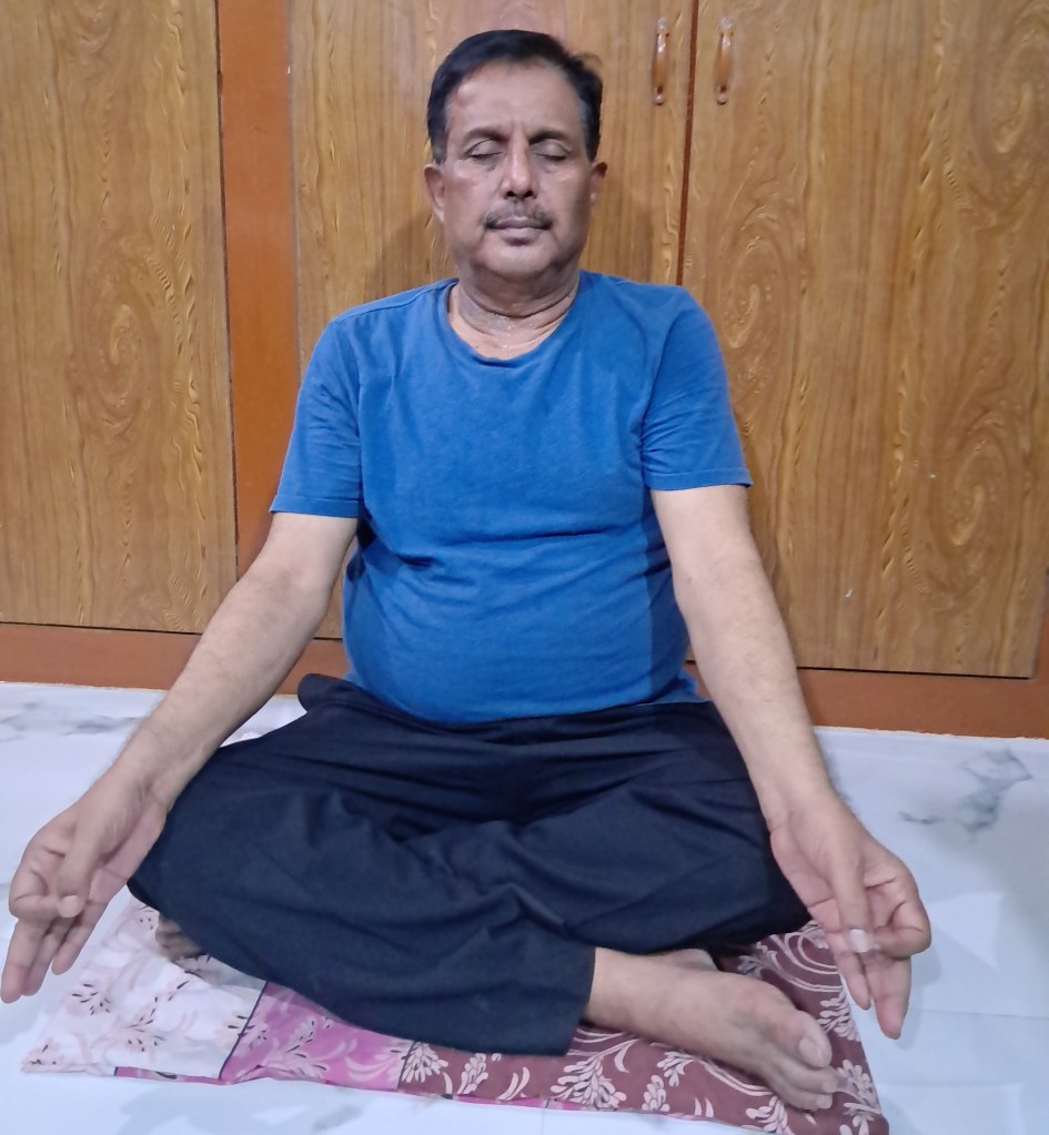
The Author Practicing Yoga

११ दिसंबर २०१४ को संयुक्त राष्ट्रसंघ में भारत के राजदूत श्री अशोक कुमार मुखर्जी ने संयुक्त राष्ट्रसंघ के आम सभा में एक एक प्रारुप पेश किया,जिसको १७७ देशों के प्रतिनिधियों ने प्रायोजित केर दिया | एवं विश्व के लगभग सभी माननीय नेताओं ने इसका समर्थन किया | 

माननीय नरेंदर मोदी जी के प्रस्ताव पर गहराई से विचार विमर्श के बाद २०१४ में ही संयुक्त राष्ट्रसंघ (यूनाइटेड  नेशन)ने एक ‘डे ऑफ़ योगा ‘ के नाम से एक प्रारुप तैयार कर ,भारत के प्रतिनिधि को सौंप दिया | 

संयुक्त राष्ट्रसंघ ने २०१५ से हर वर्ष २१ जून को  पुरे संसार में ‘अंतर्राष्ट्रीय  योग दिवस’ मनाने का आदेश पारित कर  दिया | इसके बाद से हर वर्ष २१ जून को  पुरे संसार में ‘अंतर्राष्ट्रीय  योग दिवस’ मनाया जाता है |

 पहला अंतर्राष्ट्रीय  योग दिवस कब मनाया गया ? 

वर्ष २०१५ के २१ जून को  पुरे संसार में पहला ‘अंतर्राष्ट्रीय  योग दिवस’ पुरे उत्साह एवं उमंग के साथ मनाया गया | अंतर्राष्ट्रीय  योग दिवस पे हर साल एक थीम, नारा देता है |वर्ष २०१५ का पहला थीम ‘सद्भाव एवं शांति के लिए योगा’ था | 

आयुष मंत्रालय हर वर्ष अंतर्राष्ट्रीय योग दिवस पर एक थीम, नारा देता है | 

वर्ष २०२४  का थीम है – ‘महिला सशक्तिकरण  के लिए  योग’  

वर्ष २०२३  का थीम है -‘वसुधैव कुटुम्बकम के लिए योग’,एक विश्व एक स्वास्थय|   

वर्ष २०२२  का थीम  था-‘मानवता के लिए योग’ 

वर्ष २०२१  का थीम  था- ‘स्वास्थय के लिए योग’ 

वर्ष २०२० का थीम  था -‘घर में रहकर योग करें ‘

वर्ष २०१५ में अंतर्राष्ट्रीय योग दिवस को महिमा मंडित करने के लिए भारतीय रिजर्व बैंक ने ‘अंतर्राष्ट्रीय योग दिवस’ लिखा हुआ एक दस रुपये का एक सिक्का जारी किया | 

वर्ष २०१७  में संयुक्त राष्ट्रसंघ  के पोस्टल विभाग ने योग के दस आसनों को एक पृष्ठ पर छाप कर अंतर्राष्ट्रीय योग दिवस को रेखांकित किया | (७ )

वर्ष २०१५ के २१ जून को भारतवर्ष में दिल्ली के राजपथ पर ३५९८५ लोगों के साथ माननीय नरेंदर मोदी जी एवं ८४ देशों के प्रतिनिधियों ने लगभग ३५ मिनटों के लिए योगाभ्यास किया ,जिसमे २१ आसनो को किया गया| इसके अलावा कई गावों ,कस्बों एवं शहरो में करोड़ों लोगों ने मनाया| इस के बाद से हर साल योगा में लोगों की संख्या बढ़ते जा रहें है |

अंतर्राष्ट्रीय योग दिवस मनाने का नोडल मंत्रालय कौन है ? 

भारत में आयुष मंत्रालय अंतर्राष्ट्रीय योग दिवस मनाने का नोडल मंत्रालय है | 

अंतर्राष्ट्रीय योग दिवस को मनाने के कारण ? 

 अंतर्राष्ट्रीय योग दिवस से योग के प्रचार एवं प्रसार बहुत जोर शोर से हुआ| इसके पहले योगाभ्यास होता था ,परन्तु इतना प्रसारित नही था |पहले कंही कंही  लोग योगाभ्यास करते थे| 

योग  भी  अंतर्राष्ट्रीय योग दिवस के कारण योग को कई देशों ,एवं नेताओं का समर्थन मिला ,जिसके कारण योग तेजी से बढ़ा है | श्री अरविंदो ,स्वामी विवेकानंद एवं अन्य बड़े धर्मगुरुओं ने योग को जीवन की शैली बताया है | २०१५ में पोप फ्रांसिस ने अपने प्रवचन में योग को ईश्वर प्राप्ति का रास्ता बताया|

दुनिया भर में कार्यक्रमों का आयोजन करके योग लोगों को जागरुक किया जाता है ,और इसके फायदे से सब को अवगत किया जाता है | | 

 योग से धर्म या संप्रदाय का रिश्ता ?

योग किसी धर्म या संप्रदाय का प्रचार एवं प्रसार नहीं करता है ,न ही किसी के विरुद्ध है,यह जीवन को ,मानव को उन्नति के शिखर पर ले जाने का मार्ग है ,एक प्रयास है | योग से जिन्दगी आनंद से जीने लायक हो जाता है  

योग के फायदे 

योग और ध्यान एक विषय के रुप में स्थापित हो चूका है| योग पर शोध हो रहा है| चिकत्सा विज्ञानं में  योग एक विषय है | 

योग सिर्फ शारीरिक व्यायाम नही है ,इसके साथ ही इसमें मानसिक एवं अध्यात्मिक पहलु भी है | कोविड -१९ के वक्त योग ने बहुत लोगों की जान बचाए,इसके साथ ही अवसाद  (depression)से बहुत लोगों को बचाया है |

हर दिन नियमत रुप  से  नियमत जगह पर नियमत समय तक योग करें,इससे आप  निरोग रहेंगे|योग आपको ऊर्जावान ,तंदरुस्त रखता है|  आप मानसिक शांति, भावनात्मक शांति को अनुभव करेंगे|  

पार्क में ,या किसी खुले जगह या घर के एक जगह पर योग कर  सकते है| 

अगर आप पहले से योग करते है,तो इसे नियमित रुप से करें और दूसरों को भी योग करने के लिए प्रोत्साहित करें|

अगर आप पहले से योग नहीं करते है,तो इसे नियमित रुप से योगाभ्यास करना आज से ही प्रारंभ करें | और इस वर्ष १० वा योग दिवस है,इसके लिए आप तैयारी कर ले | 

                                    आलेख पढ़ने के लिए धन्यवाद | 

आंतरिक स्रोत: https://knowledge-festival.blogspot.com/2023/06/world-environment-day.html  

https://knowledge-festival.blogspot.com/2023/06/2023international-yoga-day-2023.html

बाह्य स्त्रोत :

1.      UN Declared June 21 as International Day of Yoga Archived July 9 2016, at the Wayback Machine

2.      ^ “Yoga: Its Origin, History, and Development.” www.mea.gov.in. Retrieved June 20, 2018.

3.      ^ Jump up to a b “UN declares June 21 as ‘International Day of Yoga'”The Times of India. December 11, 2014.

4.      ^ “International Yoga Day 2021: Theme, History, Quotes, Benefits, Importance”. S A NEWS. June 19, 2020. Retrieved June 21, 2021.

5.      ^ “UN General Assembly to hold informal consultations on International Day of Yoga.” The Economic Times. October 10, 2014. Retrieved June 13, 2016.

6.      ^ 10 rupees coin of 2015 – International Day of Yoga, https://www.youtube.com/watch?v=L4oay3-JcU8&t=2s

7.      ^ “UN to issue 10 stamps of ‘asanas’ on International Yoga Day”. Business Standard India. April 19, 2017. 

अस्वीकरण:  

इस आलेख में व्यक्त किए गए विचार विभिन्न लेखों]संचार माध्यमों से लिए गए है और सभी सूचनाएँ मूल रुप से प्रस्तुत की गईं हैSaA व्यक्त किए गए विचार लेखक के निजी विचार नहीं हैं तथा इसके लिए किसी भी प्रकार से उत्तरदायी नहीं है|

Leave a comment

Healthy Kidney |Know the Health of Your Kidney

Specific parameters can help you judge whether your kidney is working correctly or not. You can also take proper action to maintain Kidney health.

Keywords: Monitor urine volume|Manage blood pressure|Stay hydrated |Healthy lifestyle|

Table of contents:

  1. Introduction:
  2. Some functions are
  3. Some tips are
  4. Takeaway

Introduction:

This article is part of my mission to provide trustworthy recent health information to support the general public, patients, and professionals globally.

This article is for everyone globally, especially medical care providers and curious people. Here, you will find human Physiology and health-related topics.

This activity aims for learners to better apply the latest scientific knowledge.

Upon completing the article, you will have increased knowledge regarding the subject and use it with great confidence.

You have two kidneys. The kidneys are vital organs like the heart, lungs, and central nervous system. They perform many essential functions for life and maintain good health. Nephrons are the functional unit of the kidney. More than one million nephrons are present in each kidney

Some functions are

1. Filtering metabolic waste products from the blood and removing it through urine.

2. Maintaining electrolyte balance.

3. Maintaining fluid balance.

4. Synthysize many hormones.

5. Maintain blood pressure.

6.Activate vitamin D.

7. Release erythropoietin to stimulate red blood cell formation.

Healthy kidneys will perform proper functions. Early detection and solving of potential issues will maintain kidney health. Kidneys send simple signals when their health starts to deteriorate. But you usually ignore the simple indications until the kidney becomes very ill.

Every year, on the second Thursday in March, World Kidney Day is celebrated to promote awareness and educate people about their wonderful kidneys. One in ten people worldwide suffers from some renal issue.

‘is celebrated One in ten people worldwide suffers from some renal issue.

Kidney diseases have an insidious onset and only become apparent when the disease becomes severe. Therefore, you might only realize your kidney needs to be fixed if you are vigilant.

Prevention is better than cure. Here are some tips to help you understand and recognize the signs of kidney health. It will guide you in maintaining your kidney health. You can prevent further deterioration of kidney health and even restore its health.

Some tips are

1. Monitor urine volume: Watch your daily urine output and become alert when it increases or decreases significantly. In both cases, there may be significant kidney problems. The average urine output is 1500 ml daily, depending on myriad factors. Keep a record of fluid intake and urine output.

2. Notice the color of your urine: Normal urine is pale yellow and clear. A color change indicates issues, and you should consult a doctor.

Dark color urine may indicate excess bilirubin in the urine

High-colored urine may indicate dehydration.

Red color urine is seen in hematuria -blood in the urine.

3. Watch haziness in your urine:

Normal urine is clear and transparent. Any haziness indicates a urinary tract infection.

4. Observe the frequency of micturition: Frequent urination may be due to urinary tract infections, metabolic diseases like diabetes mellitus, or renal stones.

5. Regularly check your blood pressure: Renal diseases are the second most common cause of high blood pressure. Some renal causes of hypertension, such as renal artery coarctation, can be cured by surgical means.

6. Keep track of your body weight: In abnormal renal functions, water retention may occur, leading to weight gain. Swelling may also occur in the dependent parts of the body, e.g., the lower limb. Check your blood sugar regularly.

7. Routine urine examinations are mandatory to detect sugar, proteins, cells, casts, sedimentation, and pH values.

8. Regular medical checkups are essential when anything appears abnormal.

Takeaway

A balanced diet, regular aerobic and anaerobic exercises, and a healthy lifestyle will protect your kidneys. Avoiding smoking and drinking will improve their health.

Monitoring kidney health is crucial for good health. Monitor the volume and color of urine, sediments, cells, and casts closely to detect renal diseases in an early stage, when they are curable.

Take the advice of a medical professional for further guidance.

Happy kidney.

Disclaimer: All possible measures have been taken to ensure the accuracy and reliability of the information; however, the author does not take any liability for the same using any information provided by the website solely to the viewers. ‘The information is provided as an educational service and public awareness. It is not medical advice. We advise you to review a reference book in case of any doubt and more accurate and advanced knowledge.

In case of any medical health issue, we advise you to seek the advice of a qualified doctor and follow his instructions.

Subscribe to get new articles

Leave a comment

Leave a comment

THANKS FOR READING

How to Estimate Serum Bilirubin by Colorimeter?

      This article will delve into the Bilirubin estimation procedure, a crucial tool in diagnosing liver diseases. Understanding this procedure is of utmost importance for medical professionals and laboratory technicians, as it can significantly enhance the accuracy of their diagnoses.

Table of content

  1. Introduction
  2. Principle of estimation:
  3. Apparatus and reagents:
  4. Procedure:
    1. To estimate direct Bilirubin:
    2. Calculation of direct Bilirubin:
  5. Procedure to determine Total Bilirubin:
    1. Calculation of total Bilirubin:
    2. Calculation of indirect Bilirubin:
  6. Quality control
  7. Precautions:
  8. Other methods to determine Bilirubin in blood:
  9. Why is bilirubin estimation performed?
    1. External link:

Introduction

Bilirubin is an orange-yellow pigment present in the blood. It is a degraded product of the hemoglobin of red blood cells. Bilirubin occurs in the normal catabolic pathway of red blood cells. The average total bilirubin level is 1.2 mg/dL of blood.

Jaundice will appear when the total bilirubin level becomes 2 mg/dL of blood and more.

There are two types of Bilirubin present in the blood. One is conjugated Bilirubin, and another is unconjugated.

Bilirubin is formed by the destruction of RBCs, mainly and usually by the destruction of senile red blood cells, but in pathological conditions, it may also be due to the destruction of normal and abnormal red blood cells.

Little Bilirubin is formed in the bone marrow during normal erythropoiesis.

After formation, Bilirubin enters the blood and liver, where a sugar molecule, glucuronic acid, is added by a process involving many enzymes called Glucuronidation. Now, this Bilirubin is water-soluble and known as conjugated Bilirubin.

Some amount of Bilirubin fails to conjugate and remains attached to albumin. This fraction of Bilirubin is water-insoluble and called unconjugated Bilirubin or indirect Bilirubin.

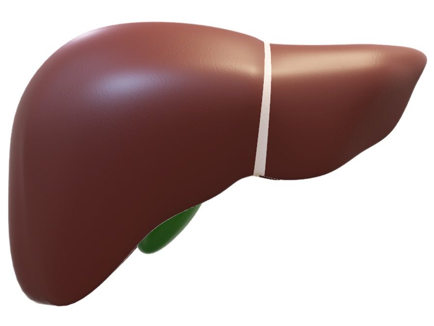
This image has an empty alt attribute; its file name is liver.jpg

LIVER

Liver

The conjugated Bilirubin is water soluble and reacts directly in aqueous solution (Direct Bilirubin).

Unconjugated Bilirubin or free Bilirubin is water-insoluble. The addition of dimethylsulphoxide makes it water-soluble and reacts (Total Bilirubin).

The unconjugated Bilirubin is determined by subtracting direct Bilirubin from total Bilirubin.

[Total bilirubin-direct bilirubin= Indirect bilirubin.]

Among the various methods for determining serum bilirubin, the Diazo Reaction stands out as the ‘Gold standard’. Its reliability and accuracy make it a cornerstone in bilirubin estimation.

Principle of estimation:

Bilirubin estimation depends on its conversion to a pink pigment known as ‘Azobilirubin’ by ‘diazotized sulfanilic acid’ and is measured photometrically using a Colorimeter. The intensity of the color developed is proportional to the bilirubin concentration in the sample.

In 1918, van den Bergh described this method for determining the bilirubin level in blood. After that, many modifications and improvements were made.

The main object is that Bilirubin reacts with diazo reagents to form pigmented Azopigments -azodipyrroles. This reaction is accelerated by the addition of some substances.

Azo pigments are measured by spectrophotometry at 530 to neutral or acid pH and 598 nm to alkaline pH.

The diazo reaction is accelerated by alcohol (methyl alcohol) and myriad compounds, such as sodium benzoate, methanol, etc. These compounds are known as accelerators. In the presence of accelerators, total Bilirubin is estimated, whereas conjugated Bilirubin is determined in the absence of accelerators.

Apparatus and reagents:

Apparatus:

Colorimeter or Spectrophotometer measuring at 555 nm.

Matched cuvettes 1.0 cm light path.

Pippete and other general pathological laboratory equipment.

Reagents:

All the agents are ready to use.

R1 Sulfanic acid 30 mmol/L
Hydrochloric acid 130 mmol/L
Dimethylsulphoxide (DMSO)7 mol/L
COMPOSITION OF REAGENT ONE.
R2Sulfanic Acid30 mmol/L
Hydrochloric acid130 mmol/L
COMPOSITION OF REAGENT TWO
R3Sodium nitrate0.74 mmol/L
COMPOSITION OF REAGENT THREE

Procedure:

To estimate direct Bilirubin:

Blood is taken from the antecubital vein, and serum is obtained by centrifuging at a speed of 3000 rpm for 30 minutes.

In a cuvette 1 cm light path, take the following reagents and serum/plasma by micropipette.

Reagents and sample Assay tube Blank tube
R2 in microliter 10001000
R 3 in microliter 50—–
dH2O —-50
Sample-serum/Plasma100100
AMOUNT OF REAGENTS AND SAMPLE in microliter

Mix and incubate precisely for 5 minutes at room temperature. Then, add the sample in the requisite amount and immediately note the reading.

Calculation of direct Bilirubin:

Direct bilirubin in mg/deciliter

=(absorbance of assay-absorbance of blank) x11.4.——CB

Where 11.4 is a standard factor, where absorbance is recorded (1).in a cuvette of 1cm path length at 550 nm wavelength, and (2) 37 degrees Celsius temperature.

Procedure to determine Total Bilirubin:

In a cuvette 1 cm light path, take the following reagents and serum/plasma by micropipette.

Reagents and sampleAssay tubeBlank tube
R 1 in microliter 10001000
R 3 in microliter 50
d H2O microliter50
Sample-serum/plasma100100
AMOUNT OF REAGENTS AND SAMPLE in microliter

Mix and incubate precisely for 5 minutes at room temperature. Then, add the sample and immediately note the reading.

Calculation of total Bilirubin:

Total bilirubin in mg/deciliter:

=(absorbance of assay-absorbance of blank) x11.4.—–TB

Where 11.4 is a standard factor, where absorbance is recorded (1).in a cuvette of 1cm path length at 550 nm wavelength, and (2) 37 degrees Celsius temperature.

Calculation of indirect Bilirubin:

Total bilirubin-conjugated bilirubin=indirect Bilirubin.

(TB-CB).

Quality control

1. Control sera are recommended to monitor the result.

2. Check the instruments, reagents calibrator, and human resources.

3. Each laboratory should have its quality control scheme and reference range.

4. There may be variations in the results of bilirubin estimation.

Precautions:

1. Serum or plasma is taken, protected from direct sunlight, and measured immediately. However, Bilirubin is stable at 2-8 degrees Celsius for four days and at—20 degrees Celsius for two months.

2. Blood sample is collected with proper antiseptic and aseptic procedures and kept properly.

2. Avoid contact with reagents.

3. Check the reagents -expiry date. Do not use expired or altered reagents.

Other methods to determine Bilirubin in blood:

1. Vandate oxidase method

2. Chromatographic methods

3. Electrophoresis methods

4. Electrochemical methods

Enzyme based

Non-enzymatic

5. spectroscopic methods

Fluorescence spectroscopy

Spectrophotometry

Luminescence spectroscopy

Direct spectrometry

6. High-performance liquid chromatography (HPLC).

7. Transcutaneous methods.

Why is bilirubin estimation performed?

The levels of both conjugated and unconjugated Bilirubin provide crucial information in the diagnosis of liver diseases such as hepatitis, cirrhosis, and jaundice. Understanding these levels is of utmost importance in the medical field.

Disclaimer: All possible measures have been taken to ensure the accuracy and reliability of the information; however, totalphysiology.com does not take any liability for using any information provided by the website solely for viewers. The information is provided as an educational service and public awareness. It is not medical advice.

If you have any medical issues, we advise you to seek the advice of a qualified doctor and follow his instructions.

https://www.bajajfinservhealth.in/articles/jaundice-symptoms

Thank You.

See you in the following article.

Leave a comment

श्री महाकाल भगवान के दर्शन यात्रा धार्मिक 

इस लेख में हम महाकालेश्वर -महाकाल के धार्मिक यात्रा एवं मंदिर के विषय में जानकारी प्राप्त करेंगे | आने वाले पीढ़ी को भारतीय त्यौहार ,उतस्वों एवं पूजा स्थलों की जानकारी देना इस लेख का मुख्य 
उद्देश्य है |
श्री महाकाल भगवान के दर्शन यात्रा धार्मिक 

Table of Contents:

  1. श्री महाकाल भगवान के दर्शन यात्रा धार्मिक 
  2. प्रस्तावना :
  3. यात्रा की तैयारी 
  4. यात्रा 
  5. दर्शन 
  6. उपसंहार 

प्रस्तावना :

कुछ दिनों से श्री महाकाल भगवान के दर्शन करने की इच्छा हो रही थी ,जो धीरे धीरे प्रवल हो रही थी | इसी बीच मेरे भतीजी का रिस्ता इंदौर में तय हुआ | १९ तारीख रविवार को रोका  तय हुआ इसमें जाना था | इसे मैने श्री महाकाल भगवान के आदेश माना और उज्जैन में श्री महाकाल भगवान के दर्शन के लिए तैयार हो गया | 

यात्रा की तैयारी 

श्री महाकाल भगवान के दर्शन के लिए बहुत लोग जाते हैं | उज्जैन जाने के लिए गया से सीधा ट्रैन  में सीट नहीं  था ,तो हमने  (मै एवं मेरी पत्नी )हवाई जहाज से जाना तय किया | ज्ञात हुआ इसमें टिकट है | १८ तारीख  को गया से दिल्ली एवं दिल्ली से इंदौर का टिकट बुक कराया , होटल में एक रुम भी बुक कराया | जरूरी समान लिए और चल पड़े  उज्जैन श्री महाकाल भगवान के दर्शन के लिए | 

यात्रा 

निर्धारित समय पर हमलोग इंदौर पहुंच कर होटल में गए | प्रातः काल के लिए एक कैब बुक कर लिए | नियत समय पर कैब आ गया | सुबह उठ कर जल्द तैयार होकर हमलोग दर्शन के लिए चल दिए | इंदौर से उज्जैन की दूरी लगभग साठ किलोमीटर है परन्तु सुबह के समय रास्ता में भीड़ नहीं रहने के कारण हम जल्द पहुंच गए | शीर्घ दर्शन के टिकट  ले  लिए ,जिसमे दर्शन में दिक्कत न हो | 

दर्शन 

मंदिर में भीड़ था ,हम लोग लाइन में लग गए और धीरे धीरे आगे बढे | जय महाकाल के उद्घोष से वातावरण उद्वेलित हो रहा था | दर्शन  के अभिलासा से उत्साह एवं उमंग से कुछ दिक्कत पता नहीं चला | ऊर्जा का संचार हो रहा था | हम लोग प्रसाद ले लिए  लाइन में धीरे धीरे आगे बढ़ कर दर्शन के स्थान पर पहुंच कर प्रसाद अर्पित किया | श्री महाकाल भगवान के दर्शन  बहुत अच्छे तरह से हो गया | मैं आत्मविभोर हो गया | लगा जीवन सफल हो गया | 

इसके बाद हम लोग धीरे धीरे मंदिर के गर्भ गृह से बहार आ गए | मंदिर परिसर में थोड़ा बैठ कर ,आराम करके मंदिर परिसर से बाहर आकर मंदिर से होटल के लिए रवाना हुए | 

होटल पहुंच कर जलपान ग्रहण किया | इसके बाद पारिवारिक कार्यक्रम मे दिन भर भाग लेने के बाद दूसरे दिन २० तारीख को इंदौर से वापस गया आ गए | 

उपसंहार 

बारह प्रसिद्ध ज्योत्रिलिंग हैं ,जिसमे महाकालेश्वर  महाकाल अद्भुत है | यह मान्यता है कि यह ज्योत्रिलिंग अपने आप उत्पन हुआ है( स्वम्भू ). | बारह प्रसिद्ध ज्योत्रिलिंग में सिर्फ यही स्वम्भू  है | 

इसका एक विशिस्ट बात है के मंदिर का मुख दक्छिन के तरफ है एवं ‘श्री रुद्र यंत्र ‘ मंदिर के गर्भ गृह के छत से उल्टा टंगा हुआ है | 

महाकाल मंदिर की स्थापना कब हुई इसके विषय में सपस्ट जानकारी नही है | परन्तु इस मंदिर का जिक्र पुराणों में है | कहा जाता है की भगवान प्रजापति ब्रह्मा ने इसे अपने हाथों से बनाया | 

उज्जैन में ईसा पूर्व तीसरे एवं  चौथे शताब्दी  के सिक्कों  पर भगवान शिव की आकृति है | ईसा पूर्व  छठे शताब्दी में  राजकुमार ‘कुमारसेन’  को राजा चंदा प्रदयता  ने  उज्जैन में महाकाल के मंदिर के नियम एवं व्यवस्था देखने को भेजा था,इसका पक्का प्रमाण है | 

कालिदास के महाकाव्य में इस मंदिर का जिक्र है | 

महाकाल देव ने हमें बुलाया ,और दर्शन देकर जीवन सफल कर दिया | उनके मर्जी के विना पता भी नही हिलता है, इतना यात्रा उनके मर्जी से ही सफल हो सका | 

अस्वीकरण :  इस आलेख में व्यक्त किए गए विचार विभिन्न लेखों]संचार माध्यमों से लिए गए है और सभी सूचनाएँ मूल रुप से प्रस्तुत की गईं है| व्यक्त किए गए विचार लेखक के निजी विचार नहीं हैं तथा इसके लिए किसी भी प्रकार से उत्तरदायी नहीं है| मानवीय भूल ,टंकण भूल भी हो सकता है इसके लिए लेखक किसी भी प्रकार से उत्तरदायी नहीं है|

Leave a comment