
Subscribe to continue reading
Subscribe to get access to the rest of this post and other subscriber-only content.
Learning will enrich your knowledge, enabling you to soar high and achieve your maximum potential in life; the sky is the limit.
Subscribe to get access to the rest of this post and other subscriber-only content.
In this article, we will explore anatomy, which encompasses various aspects of the human body and life sciences.
My mission is to provide trustworthy, recent health information to the general public, patients, and professionals worldwide.
Table of contents
Human anatomy is a branch of science that explains the structure of the human body. In human anatomy, we study the macroscopic and microscopic structures of the human body, how it is formed, and how these structures work synergistically.
The human body has long fascinated lovers, poets, scientists, physicians, and philosophers. Everyone wants to know what is inside the skin and an innocent face. Anatomy clears doubts and produces a detailed picture of the human body.
Anatomy originates from the Greek words’ ana’, meaning ‘up’, and ‘tome’, meaning a cutting. The study of anatomy involves dissecting an organism.
The study of human anatomy has been ongoing for time immemorial; however, documents indicate that the study of anatomy began approximately 2000 years ago.
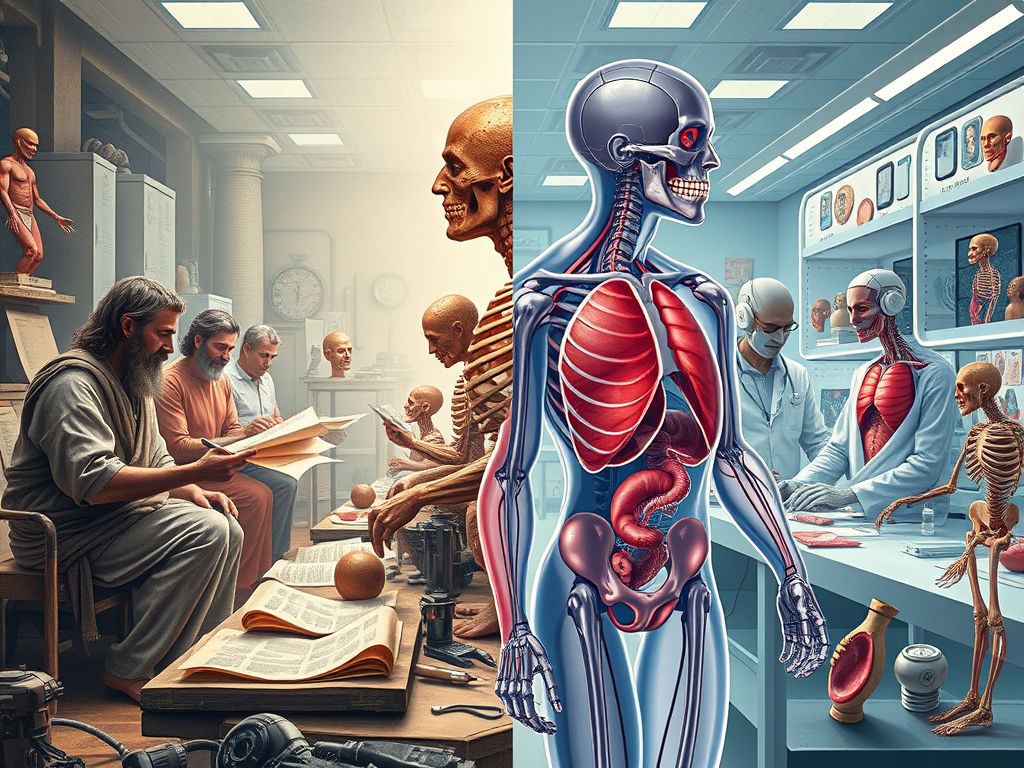
Anatomy evolved from beliefs about the human body, based on earlier texts with limited observation, to evidence-based facts, dissections, and modern digital models. This field not only underpins medical education but also plays a vital role in diagnostics, surgery, and biomedical research.
The study of anatomy is divided into
1. Gross anatomy: Studies the macroscopic structure of the body, which is divided into
Regional anatomy
Surface anatomy and
Systemic anatomy.
2. Microscopic anatomy: Examines the microscopic structure of tissues, histology, and Cytology, the study of cells.
3. Embryology –Development of the embryo.
4. Developmental anatomy: Explores the development and growth of the human body.
5. Comparative anatomy: Compares the anatomy of different species.
6. Surgical anatomy.
The study of human anatomy has been ongoing for time immemorial. Early records from Egypt (around 1600 BCE) describe anatomical knowledge from mummification.
However, it was the Greeks who first began systematic studies. Hippocrates stressed the importance of observation. Later, Galen, a Roman physician, advanced anatomical understanding by dissecting animals. His work dominated European medical thought for over a thousand years.
Herophilus of Chalcedon, with his junior Eresistraus of Ceos, performed the first systematically documented human dissection. And challenged prevailing beliefs about the human body, based on earlier texts with limited observational data. Their findings lay the foundation for the future of anatomical studies and influence the study of anatomy for centuries to come.
In India, Pandit Madhysudan Gupta (1800- 1856) performed the first human dissection at Calcutta Medical College in 1836.
The actual breakthrough came during the Renaissance. Andreas Vesalius, a Flemish anatomist, challenged long-held Galenic views. In 1543, his landmark work De humani corporis fabrica revolutionized anatomy by advocating for human dissection and the use of accurate illustrations. This period marked the birth of modern anatomy as a scientific discipline. Leonardo da Vinci and others made significant contributions to the field of anatomical illustration and understanding.
Vesalius is considered the father of anatomy.
Dissection of cadavers is the foundation of medical education, providing hands-on experience and an appreciation for human variation.
A histological study of slides reveals the microscopic structure of an organ.
Modern imaging techniques, such as ultrasound, MRI (Magnetic Resonance Imaging), and CT (Computed Tomography), have improved the teaching method of anatomy. These non-invasive tools enable the detailed visualization of internal structures in living patients, thereby enhancing diagnostics and surgical skills.
Virtual anatomy labs, 3D models, and augmented reality (AR) tools now supplement traditional learning. Platforms like the Anatomage Table and apps like Complete Anatomy provide interactive ways for students to explore the human body. Moreover, the plastination technique preserves biological tissues to offer lifelike specimens for study and display.
The future of human anatomy promises exciting developments driven by technology and interdisciplinary collaboration.
Artificial intelligence (AI) is being integrated into radiology and surgical simulation, enabling real-time anatomical recognition and predictive modelling.
3D bioprinting is another frontier. Researchers are already printing tissues and organ prototypes using living cells, potentially revolutionising transplantation and regenerative medicine. This requires detailed anatomical precision, linking anatomy directly with biotechnology.
Genomic and molecular anatomy will gain importance, as microscopic structural changes are linked to genetic expressions and diseases. Functional and systems-level anatomy will also expand, integrating physiology and anatomy for a holistic understanding of the body.
In education, fully immersive virtual reality (VR) environments may soon replace or augment cadaveric dissection, especially in regions with limited resources. Global access to high-quality anatomical education through online platforms could help bridge disparities in medical training.
1. Medical education: Anatomy is a fundamental subject in medical education.
2. Surgery: Anatomical knowledge is crucial for surgical procedures.
3. Research: Anatomy provides various fields for research.
4. Clinical practice: Understanding anatomy is essential for diagnosis and treatment.
1. Understanding human structure: A Clear concept of Anatomy provides a foundation for understanding the human body’s structure and function.
2. Informing medical practice: Anatomical knowledge informs medical practice, from diagnosis to treatment.
3. A clear concept of anatomy is essential for aspiring surgeons.
Anatomy is a primary subject of medical degrees worldwide. Many universities offer bachelor’s, master’s, and doctoral degrees in the field of anatomy.
Anatomy is an essential subject in medical education for M.B.B.S. degrees and is included in the first professional year of the M.B.B.S. curriculum.
B.Sc. Programs. M.Sc. and PhD anatomy courses are available in many universities.
An anatomist may get posted in medical colleges as a tutor, assistant, associate Professor, or Professor and dean.
An anatomist may pursue research work and may start private fitness clinics. Anatomists can work in various clinical settings, such as fitness centers, community organizations, and corporate settings.
As anatomy advances, several jobs are likely to increase in demand. Many career opportunities exist in physiology.
Promising career
An anatomist has a bright career. Anatomy has a promising future, with the advancement of science and technology, and it is developing rapidly. The human body is a highly complex system, and simulations may not fully capture its intricacies.
The scope of anatomy is broad, and its applications are diverse, making it a fundamental discipline in medicine and biology.
As we move forward, anatomy will remain a pillar of medicine, constantly reshaped by innovation, ensuring better care and deeper insight into the human form.
Thank you very much for reading. I appreciate that you took the time to spend with us. If you enjoyed reading, I’d appreciate it if you could do me a small favor. Please leave a review or a comment. Your comments will encourage other folks to read. Thanks for reading.
Thanks to AI,
Thanks to my teachers and the authors of the books I have studied, from which I have learned a great deal.
Disclaimer:
All possible measures have been taken to ensure the accuracy and reliability of the information; however, ‘learn-and-fly.co.in’ does not accept any liability for the use of the information supplied by the website by its viewers. The information is provided as an educational service and for public awareness. It is not medical advice. We recommend reviewing a reference book in case of any doubt, for more accurate and advanced knowledge.
Written by Priynka Kumari, M.Sc.(Anatomy) Facts verified by Dr. B.K. Prasad, Professor.

Subscribe to get access to the rest of this post and other subscriber-only content.
This article describes physiology, with a special focus on human physiology and its various branches. Here, learn about the past, present, future, and scope of physiology. In short, physiology refers to the study of living organisms.
My mission is to provide trustworthy, recent health information to the general public, patients, and professionals globally.
Introduction:
Physiology refers to the study of the functions of living organisms. It is divided into many branches. We are concerned with Human Physiology. In Physiology, we learn about the mechanisms that control body functions.
Physiology is the branch of biological science that deals with the functioning of living organisms. It also describes the organs that initiate and control these functions.
A French physician, Jean Fernel (1497-1558), introduced the word “physiology” for the first time.
The word “physiology” is derived from a Greek word with a Latin equivalent, “Physiologia,” which means natural knowledge. Physiology comes from two words: physis, meaning ” nature”, and logia, which means” study”.
The ancient Greek phrase” nature origin” and its study indicate an innate scientific understanding of a living body’s station, functions, and mechanisms.
Physiology is the branch of life science that deals with body functions and how these functions occur in a controlled manner. It also describes the organs that initiate and maintain these functions.
A living organism comprises different organs that respond to environmental changes to survive. Body organs function in such a way that mild variations in the external environment cannot alter the body’s internal environment, which is maintained within a normal range by a mechanism known as homeostasis.
The study of human physiology has been ongoing for time immemorial. The first authentic available record is found in the Sushruta Samhita and Charak Samhita, dating back to around 600 BCE. They proposed that vata, pitta, and kapha regulate the body’s functions.
Documents reveal that the study of medical or human physiology started at the time of Hippocrates, i.e., late 5th century BC, in Greece.
Later, many scientists, philosophers, and thinkers contributed to the development of physiology. Some of them are Galen, Aristotle, Claude Berard, Walter B. Cannon, and many others.
However, their knowledge was speculative and descriptive, lacking scientific experimental support.
A breakthrough came with Galen (129–200 AD), a Roman physician who performed animal dissections and produced detailed anatomical and physiological texts. However, many of his theories were eventually discarded with the advent of new scientific procedures and technology.
William Harvey and many others in the 17th century revolutionised physiology by describing the functions of many organs and systems supported by experiments.
A living organism is composed of many different organs that respond to changes in both its internal and external environments. Any shift in physiology will impact an individual’s physical and mental functions. The study of physiology focuses on the organs, systems, and the biological basis of the human body’s operations.
Physiological condition refers to the normal function of the body, while pathological condition refers to altered physiology, or abnormal conditions.
1. Biophysical processes
2. Biochemical processes,
3. Homeostatic control
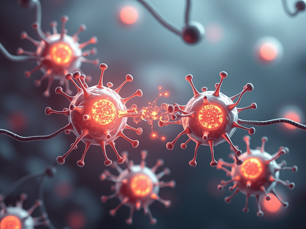
4. Rapid and precise communication between cells occurs in various ways, for example, through chemical and electrical methods.
Animal experiments have provided deep knowledge of human physiology. Physiology is a core component of medical studies worldwide, and many universities offer bachelor’s, master’s, and doctoral degrees in the field of physiology.
Modern human physiology encompasses biology, physics, and chemistry, enabling a precise understanding at the cellular, tissue, organ, and systemic levels.
Today, physiology is not limited to describing functions but explaining them through evidence-based mechanisms.
1. Cellular and Molecular Physiology:
Cellular and molecular physiology explores the roles of ion channels, neurotransmitters, hormones, and genes in regulating various bodily functions.
2. Systems Physiology:
Cells form organs, and organs form systems such as the nervous, cardiovascular, respiratory, renal, endocrine, gastrointestinal, and reproductive systems. Systems Physiology is now studied with integrated perspectives.
3. Diagnostic and Imaging Advances:
Technologies such as MRI, CT scans, PET scans, echocardiography, and EEG enable the real-time observation of physiological processes. These tools provide insights into how systems respond normally.
4. Computational Physiology:
Physiological data is now being analysed through mathematical models and simulations. This computational approach facilitates the prediction of outcomes, simulation of diseases, and testing of potential treatments, thereby reducing reliance on invasive procedures or animal models.
5. Education and Research:
In medical education, physiology is taught not only as an academic subject but as a clinical science. It forms the backbone of medical science. Modern teaching aids, including the use of simulations, virtual labs, and interactive models, have significantly enhanced learning and retention.
6. Sports physiology:
Sports physiology deals with the study of the physiological challenges associated with sports. And how to deal with them in an integrated manner.
7. Genetic counselling:
Nowadays, genetic counselling is available to prevent many hereditary diseases.
The future of human physiology lies at the intersection of advanced technologies, artificial intelligence, genomics, and personalised medicine. The coming decades promise profound transformations in how we understand and apply physiological principles.
1. Artificial Intelligence:
AI can analyse massive physiological datasets, such as ECG recordings, imaging scans, and genetic data, to identify patterns and predict health outcomes.
2. Machine learning models will assist in diagnosing conditions like arrhythmias, respiratory failure, or hormonal imbalances based on real-time physiological inputs.
3. Personalised Physiology and Genomics:
Understanding the human genome has opened the door to personalised medicine. Future physiology will not generalise bodily functions but will consider an individual’s genetic, epigenetic, and environmental factors.
4. Bioengineering and Artificial Organs:
Bioengineered tissues or artificial organs may replicate physiological functions.
Organs-on-chips and 3D bioprinting aim to mimic real physiological responses, offering potential solutions for organ failure. Research is ongoing into bionic limbs that respond to nerve impulses, replicating natural movement.
5. Devices and Remote Monitoring devices at affordable prices are available:
Smartwatches, pulse oximeters, and biosensors are used to measure heart rate, oxygen concentration, respiratory rate, and blood glucose levels. Digital blood pressure machines enable you to measure your blood pressure accurately. These machines are available at affordable prices.
Devices to monitor additional parameters, such as hormone levels, electrolyte balance, and blood pressure in real-time, are in development.
6. Space and Extreme Physiology:
As humans venture into space and colonise new environments, understanding how extreme conditions affect physiology will become crucial. Microgravity research has already revealed changes in bone density, cardiovascular function, and immunity. Future studies will investigate methods for preserving human physiology under such conditions.
7. Neurophysiology and Brain-Machine Interfaces:
Neurophysiology is rapidly advancing with the development of brain-machine interfaces. Devices like Neuralink aim to decode brain signals to control prosthetics or communicate directly with computers. Such breakthroughs could revolutionise care for patients with paralysis or neurodegenerative disorders.
Physiology is a primary subject of medical degrees worldwide. Many universities offer bachelor’s, master’s, and doctoral degrees in the field of physiology.
Physiology is an essential subject in medical education for M.B.B.S. degrees and is included in the 1st professional curriculum of M.B.B.S.
B.Sc. M.Sc. and PhD physiology courses are also available in many universities.
A physiologist may get posted in medical colleges as a tutor, assistant, associate Professor, or Professor.
Exercise physiology is the study of human physical activity and exercise. The word exercise comes from the Latin word drive forth.”
Exercise has been considered essential to human health for thousands of years in ancient cultures. The Greek physician Hippocrates recognized the importance of exercise and advocated for daily physical activity to maintain good health.
Many career opportunities exist in exercise physiology.
As physiology advances, several jobs are likely to increase in demand.
An exercise physiologist may pursue research work.
Physiologists can work in various clinical settings,
including fitness centers, community organizations, and corporate fitness.
Sports physiologists may start private fitness clinics.
A physiologist may also begin their general clinic.
Hospitals, community facilities, industries, and nursing homes may employ clinical physiologists.
A physiologist has a promising career.
Physiology has a promising future, but it faces multifaceted challenges. With the advancement of science and technology, physiology is evolving rapidly. Integrating massive datasets requires strict data privacy protocols. The human body is a highly complex system, and simulations may not fully capture its intricacies.
Ethical concerns also surround interventions like gene editing or neural implants.
A persistent quest to understand life causes the evolution of human physiology.
Physiology is a fascinating and dynamic subject that underpins translational and clinical medicine, as well as the interface between the physical and life sciences. By studying human physiology, we gain insight into how the body maintains normal health and responds to and adapts to challenges in both internal and external environments.
Today, physiology is more integrated, evidence-based, and technologically empowered than ever. It is developing very rapidly.
Looking forward, it will become even more personalised, predictive, and digital, shaping the future of healthcare and medical science.
For M.B.B.S. students and aspiring doctors, mastering physiology is not just about passing exams — it is about developing a scientific foundation that will support every future clinical decision.
Hashtags: Hippocrates # homeostasis # exercise #physiology # sports physiology# divisions #clinical# scopes #
Internal Link: https://blog.totalphysiology.com/2022/01/homeostasis external-internal.html
External Link:
1.https://en.m.wikipedia.org/wiki
2. Entin, Pauline (n.d.)”ABrie” History of ExercisePhysiolPhysiology’rn Arizona University Retrieved 2017-06-30 from
http://jan.ucc.nau.edu/pe/exs336historyVA1.htm.
3. Ivy.John L.(2007). Exercise Physiology: Brief History and Recommendations Regarding Content Requirements for the Kinesiology Major. 59:34-411.
Subscribe to get access to the rest of this post and other subscriber-only content.
Subscribe to get access to the rest of this post and other subscriber-only content.
Subscribe to get access to the rest of this post and other subscriber-only content.
Subscribe to get access to the rest of this post and other subscriber-only content.
Subscribe to get access to the rest of this post and other subscriber-only content.
Vital capacity (VC) is the air volume that can be expired by forceful effort, following maximal deepest possible inspiration.
The subject takes a deep breath of maximal inspiration and then breathes out forcefully. The volume of air expelled indicates vital capacity.
The total Tidal volume (TV), Inspiratory reserve(IRV), and Expiratory reserve volumes (ERV) make Vital capacity.
Vital capacity measures respiratory functions and indicates respiratory and overall health conditions. It indicates functions of higher brain and respiratory centers, strength of respiratory muscles, regular nerve supply to the respiratory muscles, and functions of the lungs and chest wall.
Read more of this content when you subscribe today.
Read more of this content when you subscribe today.
You cannot change your age, sex, and height, but you can improve your posture, general health, and lung and muscle conditions by simple and effective methods.
Improving your vital capacity involves strengthening your respiratory muscles and optimizing lung function. Here are some simple, free, but effective steps:
1. Maintain a normal weight.
2. Maintain hydration.
3. Ensure quality sleep for 6 to seven hours.
4. Take a balanced diet at regular intervals.
5. Avoid stress and strain.
6. Avoid pollution and ensure good quality of indoor air.
7. Quit smoking and avoid passive smoking.
8. Quit alcohol.
9. Practice deep breathing exercises, such as deep inhalations and exhalations, to expand your lungs fully and Diaphragmatic breathing, also known as belly or abdominal breathing.
10. Practice regular aerobic exercise, such as running, swimming, or cycling, and muscle-building exercises to strengthen your muscles, especially those of your back, chest, neck, and abdomen. Before starting the exercise program, follow the advice of a qualified healthcare provider.
11. Practice Yoga

12. Maintain a healthy lifestyle.
Remember, this is for informative purposes only, based on different sources of information.
Read more of this content when you subscribe today.
Read more of this content when you subscribe today.





Leave a Reply