Protect yourself from Heatstroke.

Heat stroke-symptoms, treatment, and prevention

This article discusses recent advances in the physiology of heat stroke. It will provide deep knowledge to identify the risk factors predisposing to heat stroke.

This blog will analyze the roles of endotoxins and cytokines in producing multiple organ damage in heat stroke.

We will study the incidence of hyperthermia and fever during heat stroke and the clinical biomarkers of organs during recovery.

You will learn about protective measures and treatment modules for heat stroke to live healthily in a hot climate.

Keywords:

Self care | Wellness | Awareness | Tropical | Subtropical | Heat waves| cytokines| Endotoxins ||enzymes |Hypothalamus| Thermoregulation | Sweating| Eelectrolytes | Heat cramps | Syncope | Exhaustation | Heat waves|tropical areas|cytokines|endotoxins|enzymes

Table of content

  1. Heat stroke-symptoms, treatment, and prevention
    1. Table of contents
  2. What is heat stroke?
    1. Risk factor
  3. Types of heat stroke
  4. Causes of heat stroke:
  5. Pathophysiology
  6. Investigations
  7. Differential diagnosis:
  8. Signs and symptoms of heat stroke:
  9. Treatment
  10. Prognosis
  11. Checklist
  12. Prevention
  13. What is malignant hyperpyrexia?

About’ totalphysiology.com.‘

This article is part of my mission to provide trustworthy recent health information to support the general public, patients, and professionals globally.

Here you will find human Physiology, Anatomy, and health-related topics.

What is heat stroke?

Heat stroke, also known as Sunstroke, is a severe, life-threatening condition caused by extreme heat and is the last point on a continuum of heat-related illnesses.
Heat stroke will kill if not treated properly, promptly, and vigorously. It is a medical emergency.

Heat crampsè > If not treated> Heat syncope> If not managed > it will change into>heat exhaustion >If not recognized, overlooked, and treated, heat exhaustion will turn into heat stroke.

In this condition, “core body temperature rises to 105 to 106 degrees Fahrenheit with central nervous system complications.

Heat stroke develops rapidly within 10 to 15 minutes with or without alarming features. It will develop within 10 to 15 minutes after exposure to high temperatures.

Heat stroke damages the brain and other internal organs, leading to long-term sequelae or immediate death.

Risk factor

· Age: Older people ( above 60), infants, and children below five years are more prone to develop Heat stroke.

· Sex: More typical in males

· Athletes or military personnel exercise or do heavy duty during the hot daytime.

· Manual workers, a laborer working in exposed areas with sunlight

· A person who does not drink enough water and moves on an empty stomach.

· Immunocompromised persons

· People suffer from renal, cardiac, pulmonary, and metabolic diseases like Diabetes mellitus, hypertension, and liver diseases.

· Malnourished persons may be underweight or overweight, in a poor nutritional state, and have starvation.

· Alcoholics, drug addicts

· Persons with morbidity and on drugs, for example, diuretics, sedatives, and tranquilizers.

Types of heat stroke

Depending upon the cause of heat stroke, it is divided into:

1. Classical heat stroke: This is heat stroke without apparent exertion.

2. Exertional heat stroke due to strenuous exercise.

The incidence of heat stroke is intimately related to the heat index, which is related to the environmental heat and the percentage of humidity in the atmosphere. We will learn about the heat index in detail later.

Causes of heat stroke:

· Prolonged exposure to high temperature

· High metabolic energy production due to exercise or hard work

· Dehydration

· Reduction of heat loss by the body.

Pathophysiology

Thermoregulation depends on the hypothalamus; the body gains heat from the environment and metabolic process, while heat loss is due to sweating and sweat’s evaporation from the body’s surface.

Due to excess heat of the body temperature, the regulatory mechanism fails.

The hypothalamus controls body temperature regulation and thermoregulation.

When exposed to heat hypothalamus:

· Decreases vessel myogenic tone, causing increased blood flow in the skin and excessive sweating. Evaporation of sweat causes heat loss from the body.

The hypothalamus reduces sympathetic discharge, causing dilatation of an AV anastomosis that causes increased blood flow to the skin. Therefore, heat loss occurs rapidly.

The hypothalamus directly stimulates sweat glands to secret more sweat.

When the temperature is very high, the hypothalamus fails to control or regulate thermoregulation.

Complex immunological and inflammatory components play vital roles in the production of heat stroke.

Thermo regulatory system or mechanism fails due to intense heat, and heat loss from the body ceases abruptly.

Reduced evaporation of sweat causes heat accumulation in the body.

Intense heat disturbs enzyme activities, and protein structures are disturbed.

Enzyme and protein disturbances damage vascular endothelium, increasing vascular permeability. When the permeability of the vessels increases, fluid escapes into the interstitial space, causing hemoconcentration. Blood volume decreases, reducing GFR and urine output with high-colored urine.

Decreased blood volume causes dehydration, reduces sweating, and conserves heat. After some time, the sensation of thirst will appear.

Decreasing blood volume causes hemo concentration, which causes platelet clumping and destruction. Cell destruction produces cytokines that damage tissues and organs.

Decreased blood volume, platelet clumping, and platelet destruction cause vascular occlusion, ischemia, tissue necrosis, and organ damage. This triggers a vicious cycle that causes more and more damage to cells and organs.

Necrosis of brain tissues produces immediate effects such as seizures, convulsions, and loss of consciousness.

Damage to the intestinal mucosa occurs. Intestinal mucosa becomes more permeable to water, other toxic substances, and bacteria in the gut.

Lipopolysaccharides (LPS) of Gram-negative bacteria enter into the gut and subsequently into circulation. This produces endotoxemia.

High circulatory level of LPS causes sepsis and inflammation. Endotoxins and cytokines produce necrosis in multiple organ failures and CNS dysfunction. Numerous organ failures lead to death; the sequel may persist long after being cured.

Investigations

Diagnosis of heat stroke is based on clinical diagnosis and the patient’s history. But investigations are essential to asses patients’ conditions and to exclude other diseases.

CBC, PT (prothrombin time), PTT, Blood gas analysis, Electrolyte estimation CRP C -reactive protein

Cardio and respiratory monitoring.

Differential diagnosis:

Cerebral Malaria,

Meningitis,

Neuroleptic malignant syndrome.

Complications

Seizuresà tonic or chronic

Renal failure

Convulsion

Diabetes mellitus and other metabolic endocrinal diseases due to damage of the endocrine glands conventions

Rhabdomyolysis, and

cardiopulmonary diseases.

Signs and symptoms of heat stroke:

Hyperthermia (temperature 105 degrees Fahrenheit to 106 degrees Fahrenheit, more than 40.6 degrees Centigrade) causes disorientation. Unconsciousness may occur before the development of any signs and symptoms.

In classical heat stroke -Lack of sweating, red, hot, dry skin.

During exercise-induced heat stroke, the skin is wet, damp, red, and hot.

Dizziness, lightheadedness, headache, nausea, and vomiting.

Muscle cramps, muscle weakness, staggering.

Tachycardia, fast, rapid pulse.

Rapid shallow breathing.

Reduced urine volume and high-colored urine

Delirium, coma, and death.

Treatment

If you feel that someone has had a heat stroke, call for help: dial 911, call an ambulance, and motivate others to help.

Do not hesitate to start first aid. Remember that heat stroke is always fatal without immediate medical help.

First aid :

Move the person to a cold place under a shaded space, and remove unnecessary clothes.

Wet his skin with water from a sponge or spray and fan air over the patient to speed up evaporation.

Place ice packs on the patient’s arms, groin, neck, and back.

If possible, immerse the patient in cold water or shower tub.

In Iraq, cooling with water spray was the treatment for heat stroke in 1943.

Do not use ice for the elderly, young children, and patients suffering from chronic illnesses.

In hospitals

Rapidly reduce body temperature. Put him in an air-conditioned room.

Rapidly mechanical cooling must be done to save a life.

Management of life-threatening systemic complications.

Replace electrolytes through the intravenous route. Give him fruit and vegetable juices containing electrolytes if the patient can drink. Fruit or Vegetable juices are very effective.

Monitor respiratory rate, heart rate, oxygen concentration, and pulse.

Cardiac monitoring is a must, and cardiopulmonary resuscitation may be required.

Prognosis

The risk of death is less than 5% in exercise-induced heat stroke and more than 65% in classical heat stroke.

Thousands died in the 1995 Chicago and European heat waves in 2022.

Checklist

Avoid direct sun

Avoid outdoor work and hard work,

Use a white wide-brimmed hat,

Use a thick cotton towel to protect your head, soldier, and face from sun rays when you go out the door.

No alcohol, no coffee, and no smoking.

Wear light clothes, light-colored, loose-fitting cotton clothes.

Drink excess water and fruit and vegetable juices. Sports beverages are beneficial. Drink fluids before you go outdoors.

Prevention

Prevention is better than cure.

When the heat index is high, stay in a cool place.

Avoid outdoor work and hard work,

Take rest

Use a white wide-brimmed hat,

Use a thick cotton towel to protect your head, soldier, and face from sun rays when you go out the door.

No alcohol, no coffee, and no smoking.

Wear light clothes, light-colored, loose-fitting cotton clothes.

Dark-colored urine indicates dehydration, while light-colored urine is ideal.

Try spending at least 2 hours daily in an air-conditioned environment.

To avoid direct or indirect sunlight, try to rest for at least 2 hours during peak hours of the day.

The heat index rises 10-15 points in the direct sun within minutes.

After recovery, you are more prone to heat stroke; therefore, avoid hot weather and heavy exercise.

Heat index or humiture is a combined measurement of temperature and humidity computed by adding the temperature in Fahrenheit to the relative humidity and dividing it by 2.

An increase in humidity will reduce the evaporation of sweat, reduce heat losses, and increase body temperature.

What is malignant hyperpyrexia?

Malignant hyperpyrexia is a dangerous response in susceptible persons to some medicines, especially medicine used during general anesthesia, for example, Halothane.

A rapid and sustained rise in body temperature, muscle rigidity, fever, tachycardia, and metabolic acidosis characterize malignant hyperpyrexia.

https://doi.org/10.25911/5d6cfa510ea05

Treatment is rapid cooling, drug dantrolene, and supporting treatment.

This article will be helpful for you.

Would you like to meet again? Sign up to get an email when I publish.

Question? Email me bkp337390@gmail.com

Follow me on Instagram @ bijaykprasad

@bkpdphysiology

Follow us on Twitter @ at bijaykrishnapr3

@BijayPr60250287

And don’t forget to follow me on learn and fly.co.in

FAQ:

Q1.What is malignant hypertension?

A. Malignant hyperpyrexia is a dangerous response in susceptible persons to some medicines, especially medicine used during general anesthesia, for example, Halothane.

A rapid and sustained rise in body temperature, muscle rigidity, fever, tachycardia, and metabolic acidosis characterize malignant hyperpyrexia.

Q2.What are seven tips to protect yourself from the harmful effects of heat ?

A. Avoid direct sun

Avoid outdoor work and hard work,

Use a white wide-brimmed hat,

Use a thick cotton towel to protect your head, soldier, and face from sun rays when you go out the door.

No alcohol, no coffee, and no smoking.

Wear light clothes, light-colored, loose-fitting cotton clothes.

Drink excess water and fruit and vegetable juices. Sports beverages are beneficial. Drink fluids before you go outdoors.

Protect Yourself from Cold  Weather         

Protect Yourself from Cold.

In this article, I shall discuss simple measures to reduce the effects of cold on our bodies. You will also learn about temperature regulation, features of cold exposure, and much more.

Keywords: 

Human Physiology|Thermoregulation| Wellness| Selfcare| Self care|Positivity| Dopamine |Endorphins| Bronchoconstriction| Bronchospasm| Yoga

Table of content

  1. Keywords: 
  2. Heat loss occurs by
  3. Heat gain occurs by

This article is part of my mission to provide trustworthy, recent health information to support the general public, patients, and professionals globally.

Here you will find human Physiology, Anatomy, and health-related topics.

This article is intended for an international audience of medical care providers and learners.

This activity aims for learners to better apply the latest scientific knowledge.

Introduction

Temperature changes occur worldwide. In the winter season, environmental temperatures fall. We cannot control these falls, but we can protect ourselves from the cold climate and waves.

Tissues in our bodies perform properly within a narrow body temperature range, 35.5 to 37.5 degrees Celsius. Enzymes, hormones, and other systems perform optimally when the body temperature is within this standard limit.

Our bodies maintain the core temperature within a normal range by ‘thermoregulation.’ Peripheral temperature fluctuates to some degree and affects core temperature.

The thermoregulatory system is poorly developed in infants and children, who are more prone to colds.

The thermoregulatory system is less efficient in old age than in young age. Some diseases, like diabetes mellitus and hypertension, may be present. Because blood vessels are less flexible, the elderly are more prone to colds.

Cold weather poses significant risks to older people. The mortality rate is high during cold weather in older people.

Let us know about temperature regulation in our body. Body temperature depends on heat loss and heat gain from our bodies.

Heat loss occurs by

1. Conduction –when we touch cold objects

2. Convection- exposed in a cold climate, cold wind passes after touching our bodies.

3. Radiation –temperature transfer to the environment due to temperature differences without contact.

4. Evaporation of sweat and water vapor through expiration.

Therefore, in cold climates, we must cover our bodies with cotton clothes, woolen clothes, windcheaters, etc., and remain indoors.

Baby in proper dress, in a room.

Heat gain occurs by

1. Internal source production in the body.

2. External source-heat appliances to warm the room and body.

Steps to prevent the ill effects of a cold:

1. We must stay indoors as far as possible. Our work is essential, but our lives are precious, and we must save ourselves.

2. We must use heating appliances, electrical or otherwise. We keep ourselves warm by drinking hot beverages, such as tea and coffee, and fluids with the proper amount of electrolytes.

3. We must wear adequate clothing to cover ourselves from toe to head: socks, gloves, scarves, hats, mufflers, jackets, and coats.

4. We must exercise and remain active. Exercise and physical activities cause sweating and remove waste metabolic products from our bodies.

Muscular activities produce energy –heat.

Physical activities release dopamine, endorphins, and other hormones that protect against depression.

We should perform breathing exercises, yoga, and meditation to improve our mental health.

5. We must keep ourselves well-hydrated. We must drink fluids along with electrolytes to maintain health.

6. We must eat a nutritious diet rich in vitamins, minerals, and fiber in an adequate amount to maintain normal health.

7. We must maintain personal hygiene.

8. We must not consume alcohol and must not smoke.

9. We should follow the instructions of the ‘national weather service’ and local authorities regarding cold waves and other weather conditions.

Physiology of cold exposure:

During Cold, the blood supply in peripheral parts of our bodies —fingers, toes, palms, soles, and skin —constricts to reduce heat loss. Blood flows to the central internal organs, increasing strain on the heart and raising blood pressure. It may even cause myocardial infarction.

High blood pressure may cause confusion, disorientation, cerebrovascular accidents, hemiplegia, and paralysis. These effects are pronounced in old age, malnourished persons, and persons suffering from some diseases.

In our bodies, enzymes, hormones, and tissues work efficiently at average body temperature. The regulatory mechanism tries to maintain core temperature. However, it fails in extremely low temperatures. When body temperature fails, the functions of enzymes, hormones, and tissues are affected adversely, leading to morbidity and mortality.

Cold air may cause reflex bronchoconstriction, leading to bronchospasm and breathing difficulty.

In cold climates, the oxygen supply to tissues reduces, causing hypoxia. All cells are susceptible to hypoxia, especially the brain. When the oxygen supply to the brain is compromised, confusion, disorientation, coma, and death may occur.

How to recognize the effects of Cold:

Feeling cold, shivering, numbness in fingers and toes, weakness, lethargy, confusion, disorientation, and coma.

Your social responsibilities:

Take care of your family, relatives, friends, and others.

Remain in contact with your relatives, friends, and neighbors.

Please help others.

Keep yourself warm and healthy.

Please follow me on ‘learn -and- fly.co.in’

I am more enthusiastic, and you can get new insights from my articles later.

Please submit your comments about this article. The team will work hard to evaluate the statement and make appropriate corrections. Your comments will help improve the content.

Thank you very much for reading. I appreciate that you took the time to spend with us. If you enjoyed reading, do me a small favor. Please leave a review or a comment. Your comments will encourage other folks to read. 

Hashtags: Prevention # Self-care # Wellness# Wellbeing # Health care# Human health # Human physiology# Social science

Internal Links:   https://blog.totalphysiology.com/2021/02/fever-we-must-know.html

https://blog.totalphysiology.com/2022/01/homeostasis/html

External Links: http://www.nidirect.gov.uk

FAQ:

Q1. How to protect from the cold in winter?

A. You must dress adequately, drink warm liquids frequently, and take proper, healthy food. You must remain in a warm place and avoid outdoor work.

Q.2 How do you stay healthy during the winter season?

A. By eating healthy food, drinking frequent warm drinks, and wearing proper clothing.

Q3.Safety tips for outdoor workers.

A. There are some tips for outdoor workers to stay safe and healthy in cold weather.

Dress appropriately, take frequent breaks in warm areas, and drink warm liquids. Do not take alcohol.

Disclaimer: All possible measures have been taken to ensure the accuracy and reliability of the information; however, the author does not take any liability for using any information provided by the website solely at the viewer’s discretion. ‘The information is provided as an educational service and public awareness. It is not medical advice. We recommend reviewing a reference book for any doubts and to gain more accurate and advanced knowledge.

     चैती छठ 2024 | Chaati chhaath puja 2024

प्रस्तावना : आज के व्यस्त जीवन में हम लोग अपने व्रत त्यौहार को लगभग भुला गए हैं | यह लेख खास कर युवा में अपने व्रत त्यौहार के प्रति जागरूकता लाने के लिये है | इस आलेख को पढ़ने के बाद अपने त्यौहार , महापुरूषो के प्रति जानकारी प्राप्त करने का रुचि विकसित होगा | इस लेख का यही  उद्देश है  इस लेख में हम  चैती छठ के विषय में बहुत कुछ जानेंगे |  चैती छठ कँहा कँहा होता है  इसका महत्व एवं बहुत कुछ | 

Table of contents:

चैती छठ कब से कब तक  मनाया जायेगा ?

12 अप्रैल २०२4 से चैती छठ शुरु  होगा एवं चार दिनों तक धूम धाम से मनाने के बाद १५ अप्रैल को समाप्त होगा  | 12 अप्रैल २०२4 से नहाई खाय के साथ छठ व्रत शुरु  हो जाता है | आज नहाई  खाय में कदु दाल और भात को शुद्ध रूप से   बनाया जाता है और नहाने के बाद शांत शुद्ध मन से प्राप्त -खाया जाता है | खाद्य पदार्थ अलग अलग जगहों  पे अलग अलग ढंग से बनाए जाते है |

चैती छठ किस तरह किया जाता है ?

12 अप्रैल २०२4 से नहाई खाय के साथ छठ व्रत शुरु  हो जाता है | आज नहाई  खाय में कदु दाल और भात को शुद्ध रूप से   बनाया जाता है और नहाने के बाद शांत शुद्ध मन से प्राप्त -खाया जाता है | खाद्य पदार्थ अलग अलग जगहों  पे अलग अलग ढंग से बनाए जाते है |

 दूसरे दिन खरना होता है ,जिसमे व्रती दिन में उपवास रख कर शाम को सूर्य भगवान को प्रणाम और पूजा कर अन्न ग्रहण करते है | और उसके बाद रात में कुछ व्रती सरगही करते हैं ,जबकी कुछ व्रती उसी वक्त से उपवास करते है | पूजा के तीसरे दिन शाम को अस्तगामी सूर्य भगवान को अर्घ दिया जाता है |चौथे दिन उदयमान सूर्य भगवान को अर्घ दिया जाता है और व्रत का समापन हो जाता है |

साधारणतः अर्घ स्थानीय पवित्र  नदियों पोखरों में अर्पण किया जाता है | जिसके कारण नदियों पोखरों पे बहुत भीड़ जमा हो  जाती है ,इससे व्यवस्था में दिक्त होती है ,और हादसा होने का डर रहता है  | जिससे लोग घर के छत पे या हाता में अर्घ अर्पण करतें है |   

उदयमान सूर्य भगवान को अर्घ

छठ का महापर्व साल में दो बार मनाया जाता है | एक कार्तिक छठ  है और दूसरा चैती छठ है | दोनों छठ  चार दिनों का होता है |कार्तिक छठ पूजा का विशेष महत्ब है |

चैती छठ का महा पर्व 

नवरात्रि के समय ही चार दिवसीय सूर्योपासना  चैती छठ का महा पर्व  १२ अप्रैल से प्रारंभ हो कर १५ अप्रैल के सुबह तक मनाया जायेगा | चैती छठ १२ अप्रैल से नहाय खाय से  प्रारंभ होगा ,१३ अप्रैल  को  खरने पूजन ,१४ को संध्या अर्घ एवं १५ को सुबह के अर्घ के साथ समाप्त होगा | भारतवर्ष में कई तरह के त्योहार मनाए जाते हैं, चैत्र नवरात्रि  एवं चैती छठ का त्योहार उनमें बहुत ही महत्वपूर्ण त्योहार माना जाता है | 

 चैती छठ  कँहा  मनाया जाता है?

चैती छठ का महत्ब | 

छठ  पर्व में  सूर्य भगवान एवं छठ   मैया  की  पूजा होती है | छठ पर्व  हिन्दुओ का महापर्व है | परन्तु सभी धर्म के लोग बढ़ चढ़ कर हिसा लेते है |  इस पर्व में जनमानष  स्वतः घर से लेकर घाट तक सफाई करते है ,और पुरा शहर ,कस्वा,गांव  साफ हो जाता है |  अपने सर पर प्रसाद लेकर घाट तक जाते हैं | इस पर्व में सब बरावर हो जाते हैं | आस्था का सबसे  बड़ा पर्व  है |   

   पूजा आस्था का सबसे  बड़ा पर्व

 मान्यता  एवं आस्था का सबसे  बड़ा पर्व

मान्यता है की छठ मैया सूर्य भगवान के बहन हैं और जो लोग छठ  पर्व सच्चे मन से, आस्था के साथ करते हैं तो उनके सभी  मनोकामनाए  सूर्य भगवान एवं छठ  मैया  पूरा करते हैं | 

अस्वीकरण –  

इस आलेख में व्यक्त किए गए विचार विभिन्न लेखों संचार माध्यमों से लिए गए है और सभी सूचनाएँ मूल रुप से प्रस्तुत की गईं है| इस में व्यक्त किए गए विचार लेखक के निजी विचार नहीं हैं तथा इसके लिए किसी भी प्रकार से उत्तरदायी नहीं है| मानवीय भूल ,टंकण भूल भी हो सकता है इसके लिए लेखक किसी भी प्रकार से उत्तरदायी नहीं है|

इंटरनल लिंक :https://knowledge-festival.blogspot.com/2022/04/ram-navmi-2022-date-hindi.html

                    माँ मंगला गौरी | मंदिर | पूजा 

महाविद्या  के महादेवियाँ  

https://learn-and-fly.co.in/2024/04/03/  चैत्र नवरात्रि %e0%a4%9a%e0%a5%88%e0%a4%a4%e0%a5%8d%e0%a4%b0-%e0%a4%a8%e0%a4%b5%e0%a4%b0%e0%a4%be%e0%a4%a4%e0%a5%8d%e0%a4%b0%e0%a4%bf-%e0%a5%a8%e0%a5%a6%e0%a5%a8%e0%a5%aa-%e0%a4%a4%e0%a4%be%e0%a4%b0%e0%a5%80/

  1. चैती छठ कब से कब तक  मनाया जायेगा ?
  2. चैती छठ किस तरह किया जाता है ?
  3.  चैती छठ  कँहा  मनाया जाता है?
  4. चैती छठ का महत्ब | 
  5.  मान्यता  एवं आस्था का सबसे  बड़ा पर्व
    1. अस्वीकरण –  

      Total Body Water

Total Body Water |Average| Measurements| Distribution | Body water chart

Meta description: In human physiology, body water refers to water in the human body. Water covers about 70 to 75% of Earth’s surface. It is present everywhere and in large amounts in living organisms, including humans. In humans, it accounts for about 60% of total body weight.

Keywords:Extracellular fluid | Interstitial | Intracellular fluid| Composition of Plasma | Cholesterol | Phospholipids.|

  • Introduction
    • Sources of water
    • Sources of water loss
  • Average total body water
  • Different compartment
  • Measurements
  • Calculation
  • Functions

This article is part of my mission to provide trustworthy, recent health information to support the general public, patients, and professionals globally. Here you will find information on human Physiology, Anatomy, and health-related topics. Knowledge of human physiology will help protect your health.

Introduction

This article discusses different aspects of water in the body. Learn how the body gets water and how to remove it.

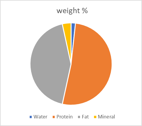
The human body has 60% water of total body weight. Protein contributes about 18%, fat 15%, and mineral body 7% of total body weight.

Our body is mostly water and distributed in different compartments. The average body fluid in adult males is about 60% water, in females, 55%, and in infants and children, about 70% of total body weight.

International Water Day: 22 March is International Water Day to recognize the significance of water in the human body and universe.

Average total body water

Total body water depends on age, sex, and adiposity.

In males -60% of body weight. In males range is 58 ± 8 %.’

In females -50-55% of body weight. In females range is 48 ± 6%.’

In obese people -45% of body weight. Because fat tissue contains only 10% of water, while muscle tissue contains 75%.

In infants -65-70% of body weight.

For example, water in a person of 60 kg is 60×60=3600/100 =36 liter. Or 36 kilograms.

Sources of water gain

Total body water –water comes into the body by:

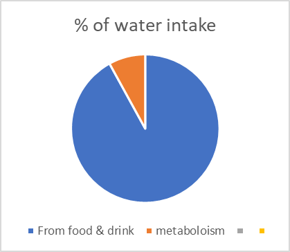
1. Liquid ingestion and water in food -2100 ml/day varies according to habits, climate, etc.

2. Formed in the body from metabolism. Due to the oxidation of carbohydrates -200 ml/day.

Total =2300 ml/day.

Chart showing % of water intake in health

Sources of water loss:

Water loss from the body

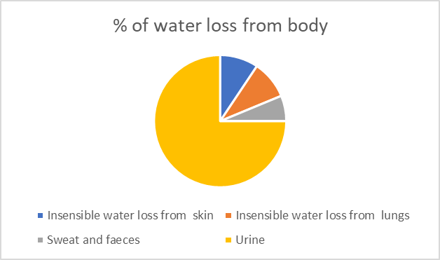
1. Insensible water loss from skin:350 ml/day

2. Insensible water loss from lungs:350 ml/day.

3. Sweat ——————————- 100ml/day.

4. Faeces——————————— 100 ml/day.

5.Urine ———————————–1400 ml/day.

     Chart showing % of water loss from the human body

The human body has 60% water of total body weight. Protein contributes about 18%, fat 15%, and mineral body 7% of total body weight. In old age and malnutrition, the total body water is less.

Water forms 45% of body weight in obese people because fat cells contain only 10% of water, while muscle tissues contain 75%.

% Contribution of water, protein, fat, and minerals in body composition

In human physiology, body water describes water present in the human body. It varies due to many factors, like age, sex, obesity, nutritional conditions, and environment.

In males, 60% of body weight.

In females, 50-55% of body weight.

In infants, 65-70% of body weight.

Detail of body water % charts and table form, showing effects of age and sex.

Sex                                Age in years
Healthy male                  Range                Average12 to 1819 to 4950  and above
52 to 66%      60%43 to 72 %      60%47 to 67%       56%
Healthy female            Range in %         Average in %   
49 to 63%41 to 60%39 to 57
56%50%47%

Total body water as % of body weight in  infants and children :

Both sexUpto six month6 months to 1 year1 to 12 years.
Range in %65 to 8555 to 6550 to 75
Average in %746060

Total body water is distributed in the following compartments :

1. Extracellular fluid 20% of body weight 14 L

a. Interstitial fluid and 11 L

b. Blood Plasma 3L.

c. Transcellular Fluid 1-2 L.

2. Intracellular fluid. about 40% 28 L

The blood volume of adults is about 80 ml/kg of body weight, which is 8% of the total body weight. The average volume is 5L.

Blood Plasma is about 3L.

Transcellular fluid is usually not taken into account in calculations. It is present in the third space—in the organs, for example, the gastrointestinal tract, the cerebrospinal space as cerebrospinal fluid, the urinary bladder, and other organs.

These data may vary depending on many factors, such as methodology and sample size.

Measurement of total body water volume

Total body water measurement is performed by

1. Flowing-afterglow mass spectrometry (FA-MS) in this method, the subject ingests a known amount of D2O (heavy water). After some time, the abundance of ‘deuterion’ is estimated in water vapor in the exhaled breath sample.

2. Indicator dilution principle: Methods based on this principle measure extracellular fluid and plasma volume.

Extracellular fluid(water) estimation: Inulin is used.

Evans Blue dye, Raio-iodinated human serum albumin (RISA), Raio-iodinated human gamma globulin, and fibrinogen are used for plasma volume measurement.

It is not possible to directly estimate intracellular water. It is estimated by subtracting extracellular fluid from total body water.

Intracellular water = Total body water- Extracellular fluid.

3. The Bioelectrical Impedance analysis method analyses total body water %. This is a non-invasive, simple, and low-cost method, and it is now widely accepted to determine total body water.

Calculation of total body water

Total body water is calculated by

TBW= body weight in kilograms x C (co-efficient factor).

The value of C is 0.6 for non-elderly males.

The value of C is 0.5 for adult females, elderly males, and malnourished males and females.

The value of C is 0.45 for elderly females.

We can calculate total body water in healthy adults using anthropometric equations. But these methods are of limited value. One such equation is Watson’s equation.

“The equations that gave the best fit were for males: total body water = 2.447 − 0.09516 A + 0.1074 height + 0.3362 weight (liters) (yr) (cm) (kg) (SD: 3.76, r2: 70.4%) and for females: total body water = −2.097 + 0.1069 height + 0.2466 weight (liters) (cm) (kg) (SD: 3.60, r2: 73.6%). Numerous other linear regression equations are present, but none are widely accepted.

https://doi.org/10.1093%2Fajcn%2F33.1.27

Functions of body water

Life is not possible without water. We can not survive without water. The functions of water in the human body are:

1. Solvent for all organic and inorganic substances in the human body.

2. Transportation of nutrients, hormones, electrolytes, and waste products of metabolism.

3. As a medium for heat loss.

4. Lubricates joints -in synovial fluid.

5.Shock absorber as in cerebrospinal fluid, pleural fluid, and pericardial fluid.

6. Water is present in the gastrointestinal tract, saliva, bile, gastric juice, and intestinal secretions.

7. Buffer action.

8. Provides viscosity.

9. Provides acid-base balance.

10. Blood coagulation.

Total body water is regulated by a homeostatic mechanism, including centers in the brain, hormones, ADH, Aldosterone, Renal mechanisms, and much more.

FAQ:

Q1. How do we calculate a total body water deficiency?

A . The following equation calculates a total body water deficiency or deficit (TBWD):

TBWD=TBW x (1- [Na+]t/ [Na+]m)

Where [Na+]t is the targeted [Na+] ion concentration, usually 140 mEq/liter, and [Na+]m is the measured concentration of [Na+].

2. How do you calculate your water percentage?

A. You can calculate your water percentage by dividing your ‘total body water’ by 60, which gives your water percentage.

3. How do I maintain a healthy water %?

A. Calculate how much water you must drink to maintain health. Weigh yourself in pounds and divide it by 2. The result in ounces will give the amount of water you must drink to maintain normal health. One pound = about 0.4535 kg, and one ounce =30 ml (approximate).

HASHTAGS: Extracellular fluid # Interstitial # Intracellular fluid# Distribution#Body water charts# Calculation# Functions #

चैत्र नवरात्रि २०२४ तारीख| Chait Navratri 2024| Hindi

प्रस्तावना : आज के व्यस्त जीवन में हम लोग अपने व्रत त्यौहार को लगभग भुला गए हैं | यह लेख खास कर युवा में अपने व्रत त्यौहार के प्रति जागरूकता लाने के लिये है | इस आलेख को पढ़ने के बाद अपने त्यौहार , महापुरूषो के प्रति जानकारी प्राप्त करने का रुचि विकसित होगा | इस लेख का यही  उद्देश है  इस लेख में हम चैत्र नवरात्रि के विषय में बहुत कुछ जानेंगे | चैत्र नवरात्रि कँहा कँहा होता है  इसका महत्व एवं बहुत कुछ | 

Keywords :  व्रत |त्यौहार कन्या पूजन |कन्या भोज | कलश स्थापित  | माँ के रूप | चार नवरात्रि | गुप्त नवरात्रि | 

Table of contents

1. चैत्र नवरात्रि  कब मनाया जाता है

2.चैत्र नवरात्रि कँहा  मनाया जाता है

3.चैत्र नवरात्रि  का इतिहास

4.चैत्र नवरात्रि  कई नाम

5.चैत्र नवरात्रि  के पौराणिक कथा

6.चैत्र नवरात्रि   का महत्व 

चैत्र नवरात्रि  कब मनाया जाता है?

Photo by the author

भारतवर्ष में कई तरह के त्योहार मनाए जाते हैं, चैत्र नवरात्रि का त्योहार उनमें बहुत ही महत्वपूर्ण त्योहार माना जाता है | यह चैत्र माह के शुक्ल  पक्छ से प्रारंभ होकर रामनवी तक चलती है |  भारतवर्ष में कई तरह के त्योहार मनाए जाते हैं, चैत्र नवरात्रि का त्योहार उनमें बहुत ही महत्वपूर्ण त्योहार माना जाता है | 

कलश स्थापना का मुहूर्त ९ अप्रैल को सुबह ७ बज कर ०२ मिनट से १० बज कर १६ मिनट है | इसी दिन – ९ अप्रैल को सर्वार्थ सिद्धि  योग एवं अमृत सिद्धि योग का संयोग है जो सुबह ७ बज कर ३२ मिनट से शाम ५  बज कर ६ मिनट  तक है | 

९ अप्रैल को दोपहर ११ बजकर ३३ मिनट से १२ बज कर २४ मिनट तक अभिजीत मुहूर्त है ,जो किसी शुभ कार्य का शुभारम्भ करने के उपयुक्त है | अगर आप कोई नया काम शुरु करने जा रहे है तो यह सही समय है | 

विद्वानों के अनुसार इस वर्ष नवरात मंगलवार से शुरु हो रहा है ,जिसके कारन माँ दुर्गा का आगमन घोड़े की सवारी से है ,जो अशुभ माना जाता है | परन्तु माँ दयालु है ,९ अप्रैल को पुरे दिन पूजा करने के बाद छमा याचना करने से शुभ फल देंगी | 

१७   अप्रैल २०२४ को रामनवमी मनाया जायेगा| रामनवमी पूजा का ९ अप्रैल को  पूर्वाह्न ११ बजकर १७ मिनट से अपराह्न एक बजकर पैतीस मिनट तक होगा |

चैती छठ का महा पर्व 

नवरात्रि के समय ही चार दिवसीय सूर्योपासना  चैती छठ का महा पर्व  १२ अप्रैल से प्रारंभ हो कर १५ अप्रैल के सुबह तक मनाया जायेगा | चैती छठ १२ अप्रैल से नहाय खाय से  प्रारंभ होगा ,१३ अप्रैल  को  खरने पूजन ,१४ को संध्या अर्घ एवं १५ को सुबह के अर्घ के साथ समाप्त होगा | भारतवर्ष में कई तरह के त्योहार मनाए जाते हैं, चैत्र नवरात्रि  एवं चैती छठ का त्योहार उनमें बहुत ही महत्वपूर्ण त्योहार माना जाता है | 

गर्मी के मौसम  शुरुआत  के साथ ही चैत्र नवरात्रि मनाया जाता है | यह चैत्र माह के शुक्ल  पक्छ से प्रारंभ होकर रामनवी तक चलती है |  भारतवर्ष में कई तरह के त्योहार मनाए जाते हैं, चैत्र नवरात्रि  का त्योहार उनमें बहुत ही महत्वपूर्ण त्योहार माना जाता है | 

 चैत्र नवरात्रि कँहा  मनाया जाता है?

चैत्र नवरात्रि भारत वर्ष का एक बहुत महत्वपूर्ण  पर्व है,जिसे पुरे भारत वर्ष में मनाया जाता है | कुछ राज्यों में जैसे बिहार ,उतर प्रदेश ,असम, झारखण्ड और पश्चिम बंगाल में बहुत उत्साह से मनाया जाता है | अन्य राज्यों में भी इसे मनाया जाता है | इसके आलावा चैत्र नवरात्रि अन्य देशों जैसे अमेरिका ,कनाडा,इंग्लैंड में भी मनाया जाता है |

 चैत्र हिंदी कैलेंडर के अनुसार पहला महीना है | चैत्र नवरात्रि नए वर्ष के पहले दिन से शुरू होता है | इस दिन हिन्दू धर्म के अनुसार नए वर्ष के पहले दिन के रूप में मनाया जाता है | गुजरात में नवरात्रि  डांडिया और गरबा के रुप में पुरे उत्साह से  मनाया जाता है |भारत के कई प्रदेश में नवरात्रि का पहला दिन ,नए वर्ष के पहले दिन के रूप में मनाया जाता है | महाराष्ट्र में यह   ‘गुरु पर्व ‘के रूप में ,कश्मीर में ‘नवरेश ‘ के रूप में मनाया जाता है | 

                                                    चैत्र नवरात्रि  कई नाम

नवरात्रि के अन्य नाम : नराते ,नवरात इत्यादि हैं | 

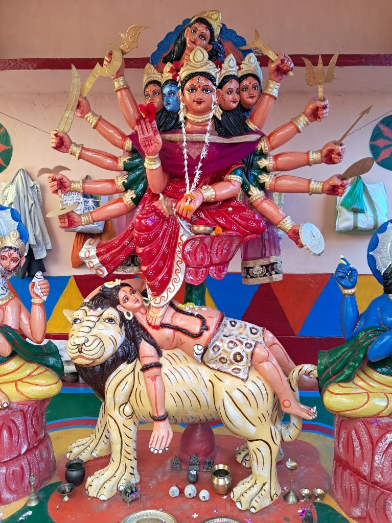
नवरात्रि एक संस्कृत शब्द है जिसका अर्थ है नौ रातें | इन दिनों माँ दुर्गा  शक्ति रुप के नौ रूपों में पूजा की जाती है एवं दसवां दिन दशहरा मनाया जाता है | चैत्र नवरात्रि में दसवां दिन रामनवमीं के रुप में मनाया जाता है | इस दिन भगवान राम का  जन्म हुआ था | भगवान राम को , विष्णु भगवान  का  सातवें अवतार  मन जाता है | 

नवरात्रि वर्ष  में चार बार में मनाया जाता है | एक नवरात्रि चैत्र में ,दूसरा पौष  में ,तीसरा असाढ और चौथा  माघ माह नवरात्रि में मनाया जाता है | असाढ और माघ माह में ‘गुप्त नवरात्रि  मनाया जाता है | इसमें चैत्र नवरात्रि एवं शारदीय नवरात्रि बहुत महत्वपूर्ण  पर्व है। 

   चैत्र नवरात्रि  के पौराणिक कथा 

भारत में हर त्योहार के पीछे कोई न कोई पुरानी कहानियां होती हैं | उसी के अनुसार भारत वर्ष में त्योहार मनाया जाता है | चैत्र नवरात्रि वैदिक युग से ही मनाया जाता है |   

एक पौराणिक कथा चैत्र नवरात्रि  के विषय में है की, भगवान राम ने चैत्र माह में माँ दुर्गा  की उपासना कर शक्ति प्रताप किया और रावण पर विजय प्राप्त किया था ,इसी  के उप्लक्छ में  चैत्र नवरात्रि मनाया जाता है | ऐसा विश्वास किया जाता है कि नवरात्रि के समय माँ  पृथ्वी पर निवास करती हैं ,अपने भक्तों के बीच रहती हैं | इससे माँ  जल्दी प्रसन्न होती हैं | 

   चैत्र नवरात्रि में भी शरद चैत्र नवरात्रि के तरह माँ भगवती माँ दुर्गा के नौ रूपों की उपासना आराधना की जाती है | 

पहला दिन माँ शैलपुत्री | 

दूसरा दिन माँ ब्रह्मचारिणी

तीसरा दिन माँ चंद्रघंटा 

चौथा दिन माँ कुष्मांडा  

पांचवा दिन माँ स्कंदमाता 

छठा दिन माँ कात्यानी 

सातवां दिन माँ कालरात्रि 

आठवां दिन माँ महागौरी 

नौवां दिन माँ सिद्धदात्री | 

चैत्र नवरात्रि का महत्व 

वैसे तो माँ की साधना किसी भी  वक्त करने से माँ  प्रसन्न होकर आशिर्बाद देती हैं | परन्तु माँ की साधना खास कर अष्ट्मी एवं नौवमीं को करने से माँ  प्रसन्न होकर आशिर्बाद देती हैं | इन दिनों पूजा का ख़ास महत्ता है |अष्ट्मी एवं नौवमीं को कन्या पूजन जिसे कन्या भोज का बहुत महत्व है | बहुत जगह पर कन्या पूजन का आयोजन किया जाता है | इसमें नौ बाल  कन्याओं (जो माँ के रूपों का दर्शाती हैं ) का पूजन किया जाता है |  

नवरात्रि किस तरह मनाते हैं ?

नवरात्रि  के पहले दिन कलश स्थापित कर के पूजा शुरु किया जाता है ,बाद में  माँ  की मूर्ति स्थापित किया जाता है | कुछ लोग घर पर कलश स्थापित कर के पूजा शुरु करते  है | पूजा  पंडाल में  माँ  की मूर्ति स्थापित किया जाता है,छठा दिन भक्तों के लिए पंडाल खोला जाता है | भक्तों की भीड़ माँ के दर्शन के लिए उमड़ परती है | दसवें दिन मूर्ति को प्रवाहित किया जाता है |  

 Hashtags: कन्या पूजन #कन्या भोज#कलश स्थापित# ,माँ के रूप#चार नवरात्रि #गुप्त नवरात्रि#अमृत सिद्धि योग #सर्वार्थ सिद्धि #उदया तिथि#सनातन धर्म# 

इन्हें भी देखें | https://knowledge-festival.blogspot.com/2021/10/maa-durga.html

अस्वीकरण –  इस आलेख में व्यक्त किए गए विचार विभिन्न लेखों]संचार माध्यमों से लिए गए है और सभी सूचनाएँ मूल रुप से प्रस्तुत की गईं है| इस में व्यक्त किए गए विचार लेखक के निजी विचार नहीं हैं तथा इसके लिए किसी भी प्रकार से उत्तरदायी नहीं है| मानवीय भूल ,टंकण भूल भी हो सकता है इसके लिए लेखक किसी भी प्रकार से उत्तरदायी नहीं है|

  Liver Donation                                                   


Liver Donation

This article is intended for an international audience of medical care providers and learners.

This activity aims for learners to better apply the latest scientific knowledge.

Upon completing the article, you will have increased knowledge regarding the subject and use it with great confidence.

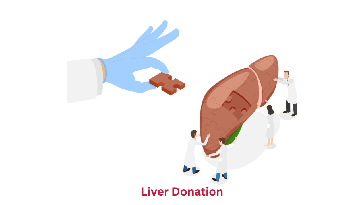
Liver donation is a pious act of charity. If you donate your liver to someone, you are prolonging his life. You are bestowing life on him.

It is unlike kidney donation, where one whole kidney is removed and donated. In liver donation, only some part of your liver is removed.

In the case of kidneys, dialysis is there to support the patient’s life, but in the case of livers, no such procedure is available to date.

The liver performs more than 500 functions and is unique in its ability to regenerate rapidly. Even when 75% of the liver is removed, it proliferates, attains its standard size, and functions properly.

Donating your liver ( part of it) has no adverse effect on your health, as your liver will regenerate rapidly and function properly.

Living liver donation is a major surgical procedure and carries all complications of surgery as well as that of anesthesia.

Now, liver donation is a very safe operation in specially designated hospitals that provide state-of-the-art techniques. Before donation, experts perform meticulous physical and mental examinations. Counseling is done. A thorough evaluation of your health is performed and certified.

Mortality is only 0.1% and morbidity is 30%.

1. Complications of general surgery and anesthesia.

2. Bleeding

3. Blood clot formation

4. Wound infection

5. Wound dehiscence

6. Bile leaks

7. Hernia

8. Pain and discomfort at the site of incision.

9. Nausea and vomiting.

10. Psychiatric problems may develop anxiety, depression, etc.

11. In some cases, the liver may fail to regenerate or work properly after the removal of a portion of the liver -it may become fatal.

A seven-day hospital stay is mandatory.

It takes about three months for a full recovery.

Disclaimer: All possible measures have been taken to ensure the accuracy and reliability of the information; however, ‘totalphysiology.com’ does not take any liability for using any information provided by the website solely to the viewers. ‘The information is provided as an educational service and public awareness. It is not medical advice. We advise you to review a reference book in case of any doubt and more accurate and advanced knowledge.

If you have any medical issues, we advise you to seek the advice of a qualified doctor and follow his instructions.

                      Who can Donate Blood in India?

H1 Who is eligible for Blood Donation in India?

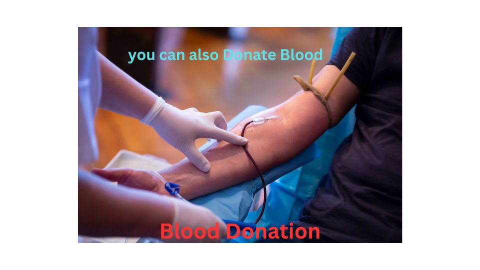
Adequate blood is vital to life. Without sufficient blood, we cannot survive. Blood is formed in the body; despite tremendous development, we cannot manufacture blood outside our bodies. Therefore, blood donation is of paramount importance to save human lives.

National Voluntary Blood Donation Day is 1st October

H2 Table of contents

Who can donate blood in India?

Criteria of Blood donation?

Who can donate blood in India?

Who can not donate blood in India?

Are there any health benefits for regular blood donors?

Side effects on your health?

Who can donate blood in India?

Criteria of Blood donation?

Let us explain the criteria in detail.

Who can not donate blood in India?

Are there any health benefits for regular blood donors?

When you donate blood, you have some mental and emotional benefits. The Mental Health Foundation reports that giving blood reduces stress and improves emotional well-being. You feel your blood will save at least three lives, leading to happiness in their families. You feel exalted. Some societies will encourage you. Last but not least is your faith that you will get blood in an emergency.

There is no proven beneficial effect on your physical health.

Side effects on your health.

Blood donation is safe. There is no risk of infection, as new sterile equipment is used for each donor, and trained health professionals are present during donation with all life-saving measures.

However, you may feel dizziness, weakness, and pain at the venepuncture site.

FAQ

Can a woman donate blood during pregnancy?

No.

Can a woman donate blood during menstruation?

No.

Can a 17-year-old boy donate blood in India?

In India, he can not donate blood.

Can a 67- year-old donate blood in India?

In India, he can not donate.

How much blood is donated at a time?

About 350 ML.

How much blood is present in one unit?

350 ML

Links:

http://nbtc.naco.gov.in/page/eligibility/%22

https://www.who.int/campaigns/world-blood-donor-day/2018/who-can-give-blood

https://www.google.com/url?sa=t&source=web&rct=j&opi=89978449&url=https://asbtc.assam.gov.in/frontimpotentdata/eligibility-criteria-for-blood-donation&ved=2ahUKEwjf4f_OlJmFAxX8hGMGHYA9DgoQFnoECBEQBQ&usg=AOvVaw1eOOQuGYFj2xfOZGViLmaU

What Is Obesity? Learn the Causes and Effects Of Obesity in Detail

How is obesity defined? What is its prevalence? Let us delve into its causes and effects and how we can eliminate obesity. We will also try to take measures to prevent it. 

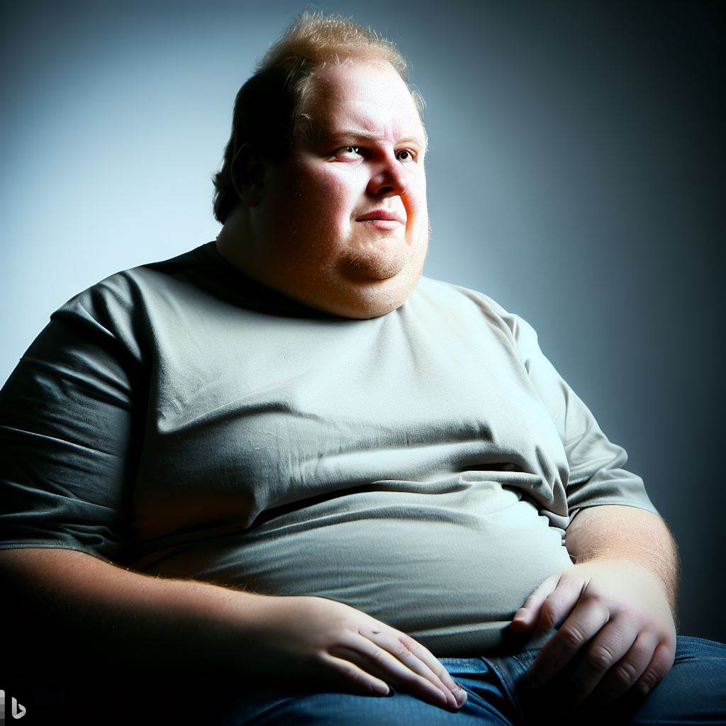
Definition: Obesity is the abnormal and excessive fat accumulation in the body. It is a chronic disease associated with other chronic diseases. The word obesity originates from the Latin ‘Obesitas, ‘ meaning fat, stout, or plump. Obesity was known in the early historical period. 

Obesity is related to various conditions and syndromes, especially with Diabetic type 2, osteoarthritis, cardiovascular diseases, hypertension, obstructive sleep apnea, asthma, some cancers, etc.

Incidence: It is more common in females and is prevalent in adults as well as in children. Globally, 650 million people are obese, which is rising rapidly.

Diagnosis: The diagnosis of obesity is based on the Body Mass Index (BMI) and is further evaluated in terms of the Waist-Hip ratio, waist-to-height ratio, and total cardiovascular risk factors.

Category                     BMI Kg/meter square

Underweight                    >̲18.5

Normal weight                   19.5–24.9

Overweight                        25—29.9

Obese class I                      30-34.5

Obese class II                     35-39.9

Obese class III                     >40

Some countries use lower values. For example, in Japan, BMI >25 is considered obese, while in China, it is >28.

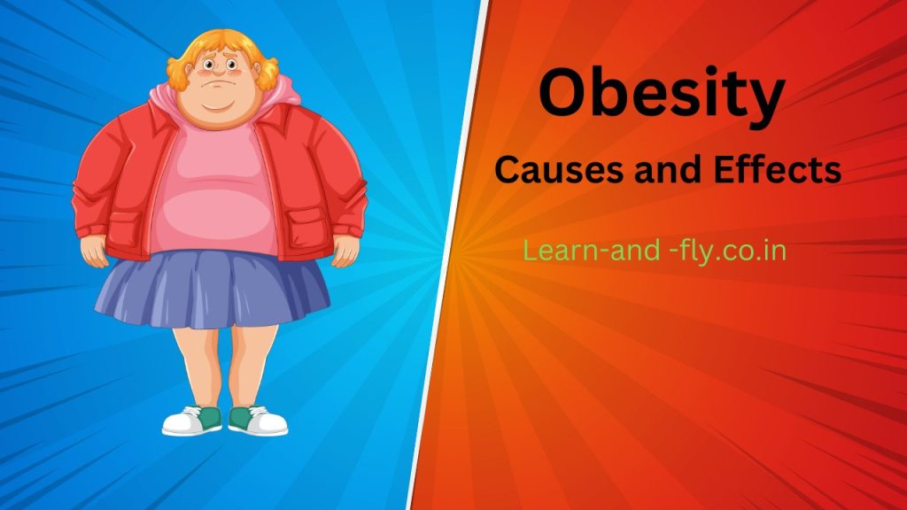
AN OBESE FEMALE FIGURE BY CANVA-

Causes: Obesity is due to 

1. Persistent positive energy balance is responsible for obesity.

2. The body weight set point is raised.

3. Genetic factors: Alterations in the various genes controlling appetite and metabolism predispose to obesity. Fat mass and obesity-associated genes (FTO gene ) are detected. Many hypotheses exist, but none are widely accepted.

Obesity shows inheritance. If both parents are obese, 80% of children will be obese. When both parents are normal, children are only 10% likely to be obese.

It is associated with rare genetic syndromes like Prader-Willi, Bardet-Bedi, Crohn’s, and MOMO.

4. Individual causes are -diet habits, physical activity, and sedentary lifestyle.

A calorie model- excess food intake and lack of physical activity.

5. Eating disorders like ‘binge eating syndrome ‘and night eating syndrome ‘are also responsible for obesity.

Carbohydrate insulin model –High glycemic load causes hormonal changes that promote caloric deposits in adipose tissue.

6. Socioeconomic factors,

Malnutrition in early life causes endocrinal changes, which may promote obesity later.

7. Environmental factors

8. Endocrinal factors: Hypothyroidism, Cushing syndrome, Growth hormone deficiency, etc.

Gut flora also plays a vital role in the development of obesity.

9. Drugs: Insulin, sulfonylurea, steroids, phenytoin contraceptives, and many more.

Obesity increases the risk of clinical depression, and depression increases the chance of developing obesity.

The complications of obesity:

Complications of obesity were well known. The great surgeon of India, Sushruta, had reported about obesity. He related it to heart disease and mentioned that physical activity would cure it.

Obesity directly :
Increases fat mass
Causes osteoarthritis,
Non-alcoholic fatty liver,
Alters the body’s response to Insulin.
Cardiovascular disease,
Hypertension,
Lung diseases,
Mental disorders, depression, anxiety and stress.
Gout,
Low back pain,
Multiple sclerosis,
Carpal tunnel syndrome, etc.
Pregnancy-related complications:

Congenital disabilities

Intrauterine fetal death,

Infertility,

Pregnancy and delivery complications, etc.

In certain conditions, obesity is helpful –positive effect, e.g., in heart failure or hemodialysis. This is the ‘Obesity survival paradox.’

Prevention of obesity:

Lifestyle change-give up smoking and alcoholic drinks.

A healthy and timely diet rich in dietary fiber.

Reduce intake of high fat and high carbohydrate diet

Start an active life.

Regular exercise.

An Introduction to Photoreceptors of the Human Eye

 Introduction

Rods and cones are photoreceptors present in the -retina. Each rod and cone are divided into 1.Outer segment 2.Inner segment, and 3.Synaptic zone.

  1.  Introduction
  2. The outer segments:
  3. Regularly placed piles of flattened discs or saccules of the cell membrane are present in the outer segments of rods and cones. Usually, discs are present in the rods and saccules in the cones. The discs and saccules contain photosensitive pigments.
  4. Photosensitive pigments
  5. The inner segments:
  6. Mitochondria are abundant in the inner segments of rods and cones. The inner segments of rods are thin, but those of cones are thick and large.
  7. Structure and functions of photosensitive pigments
  8. Regeneration of rhodopsin:
  9. The photosensitive pigments are
    1. “Sensitivity range of vision” or “Visibility range of vision”
    2. Types of vision:
      1. Photopic vision: It is daylight vision due to cones. Cones are stimulated at higher intensity of light -about one millilambert (1m A.)
      2. Scotopic or dim light vision is due to rods, and get stimulated below 0.001 m A. Rods are extremely sensitive to light.
      3. Mesopic vision is full moonlight vision, and the brightness level is 0.01m A.
    3. Disclaimer: All possible measures have been taken to ensure the accuracy and reliability of the information; however, ‘totalphysiology.com’ does not take any liability for using any information provided by the website solely to the viewers. ‘The information is provided as an educational service and public awareness. It is not medical advice. We advise you to review a reference book in case of any doubt and more accurate and advanced knowledge.
    4. Subscriber Content
    5. Subscribe to get access

The outer segments:

Regularly placed piles of flattened discs or saccules of the cell membrane are present in the outer segments of rods and cones. Usually, discs are present in the rods and saccules in the cones. The discs and saccules contain photosensitive pigments.

In the rods, the discs are separated from the cell membrane. However, saccules in the cones are formed by the infolding of the cell membrane.

The outer segment of the rod is a thin rod-like structure, while that of the cone is conical.

Photosensitive pigments

The photosensitive pigment in the rod is rhodopsin, which is very light-sensitive. This rhodopsin is responsible for dim light vision, i.e., scotopic vision.

In a cone, the photosensitive pigment is idopsin, responsible for bright light vision, photopic vision, color, and high visual acuity.

The inner segments:

Mitochondria are abundant in the inner segments of rods and cones. The inner segments of rods are thin, but those of cones are thick and large.

Rods are abundant in the human eye. It is estimated that about 120 million and 6 million cones are in each eye.

Rods are abundant in the peripheral parts of the retina and absent in the fovea. Fovea has only cones.

Structure and functions of photosensitive pigments

In rods, the photosensitive pigment is rhodopsin, which is present in the discs. It is coupled to the G-protein and is known as Visual purple.it is purple-red and stimulated by blue-green light with a wavelength of 505nm.

In cones, the photosensitive pigment is idopsin in the saccules. Structures of rhodopsin and idopsin are the same, except the latter has “photopsin” instead of “scotopin.”

The rhodopsin is composed of 1.retinene1 and scotopin.

Retinene1 is an aldehyde of vitaminA1.

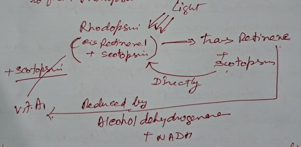
In the dark, retinene is in the 11 cis-form. When light strikes, rhodopsin 11 cis-form is converted into trans-form, and scotopic is released. Rhodopsin gets bleached.

Regeneration of rhodopsin:

1. Some bleached rhodopsin recover directly.

2. The ‘alcohol dehydrogenase’ enzyme reduces the majority of trans-retinene in the presence of NADH to vitamin A1. This, in turn, combines with scotopin to form rhodopsin.

Cones are divided into three types according to the presence of pigments in their saccules.

The photosensitive pigments are

1. Cyanolabe responds maximally to blue light, with a wavelength of 430 nm.

2. Chlorolabe – responds maximally to green light, with a wavelength of 535 nm.

3. Erythrolabe- responds maximally to red light, with a wavelength of 575 nm.

These three pigments are responsible for the primary colors: blue, green, and red. Other colors are a mixture of these colors in different proportions.

“Sensitivity range of vision” or “Visibility range of vision”

Humans can see light of wavelength between 400 nm to 750 nm.

The choroid layer of the retina absorbs ultraviolet light (wavelength of less than 400 nm).

Infrared light has a wavelength above 750 nm, and the cornea absorbs it.

The visual threshold is the minimum amount of light that produces a sensation of light.

Effect of light on rhodopsin and its regeneration.

Types of vision:

Photopic vision: It is daylight vision due to cones. Cones are stimulated at higher intensity of light -about one millilambert (1m A.)

Cones are responsible for bright light vision, color vision, and excellent visual acuity.

Scotopic or dim light vision is due to rods, and get stimulated below 0.001 m A. Rods are extremely sensitive to light.

Between 0.001 to 1 m. A is a transitional zone called the “Duplicity Theory of Vision.”

Mesopic vision is full moonlight vision, and the brightness level is 0.01m A.

Below 0.1 m A, the reading is complex, and prolonged exposure to light 100000 m A will damage the retina.

Disclaimer: All possible measures have been taken to ensure the accuracy and reliability of the information; however, ‘totalphysiology.com’ does not take any liability for using any information provided by the website solely to the viewers. ‘The information is provided as an educational service and public awareness. It is not medical advice. We advise you to review a reference book in case of any doubt and more accurate and advanced knowledge.

Subscribe to get access

Read more of this content when you subscribe today.

Hearing loss and tests

In this article, we will learn about the definition of deafness or hearing loss. We will discuss types of hearing loss, their causes, and much more.

Table of contents:

1. Introduction

2. Types

3. Causes

4. Hearing tests

5. Treatment

Hearing impairment is the inability to hear or the reduction in the hearing capacity of an average person.

Sound waves reach the cochlea in two ways:

1. Through air in the external and middle ear ( air conduction). Sound waves produce movements in the tympanic membrane, which initiates movements in the ossicles present in the middle ear. Movements in the ossicles of the middle ear cause vibration in the basilar membrane.

2.Through skull bones( bony conduction.) Sound waves produce vibrations in the skull bones, including the cochlea, which triggers vibrations in the basilar membrane.

Types:

1. Conductive deafness

2. Neural deafness or sensorineural deafness, and

3. Mixed deafness.

In conductive deafness, there are defects or impairment of functions in the conduction of sound waves. It usually causes incomplete deafness.

In Neural deafness, there may be defects in the conversion of sound waves into action potential and its transmission through the auditory pathway. Neural deafness is also known as ‘central deafness.’ It causes complete deafness.

Mixed deafness is due to causes of conductive and neural deafness.

Common causes of conductive deafness defect in :

1. External ear block in external auditory meatus by wax, foreign body, atresia, or tumor of the external auditory canal.

2. Middle ear:

  • Tympanic membrane perforation of the tympanic membrane, thickening of the tympanic membrane.
  • Infection of middle ear-otitis media.
  • Ossicles- impaired movements in ossicles due to infection.
  • Otosclerosis.
  • Eustachian tube obstruction is usually due to infections.

Common causes of Neural deafness:

1. structure of cochlea

  • Degenerative changes due to ageing-senile degeneration
  • Toxic degeneration of hair cells -drugs,
  • Prolonged exposure to loud sounds.

2. Tumors in the neural pathway or adjacent area compress the pathway.

3. inflammation of the labyrinth- labyrinthitis.

4. Miniere’s disease

5. Trauma of skull.

6. Genetic mutations in some proteins like Connexin 26, Myosin-VI, Myosin-VIIa, Sulfate transport protein, and Barttin.

Genetic mutations will cause mixed deafness.

Hearing tests:

The subject sits on a comfortable chair. The examiner explains the procedure to the subject.

1. Examination of external ears

It starts by examining the external ears, the pinna, and the external auditory meatus.

The pinna is examined for its structure and evidence of any physical abnormalities.

The examiner examines the external auditory meatus. The examiner stands in such a way that he can observe inside the external auditory meatus. The pinna is pulled gently upward and backward to straighten the external auditory meatus.

Points of observation: wax, foreign bodies, and any growth.

Now, an ear speculum is introduced gently in the external auditory meatus. A beam of light is thrown into the ear speculum to visualize the tympanic membrane. The tympanic membrane becomes visible and examined in different directions and positions.

The tympanic membrane is recognized by its oval shape, concavity pointing medially, cone of light, and glistening, shining look.

The center of the tympanic membrane appears depressed-this is the umbo.

The upper part of the tympanic membrane is pinkish and appears flaccid, while the lower and significant part appears blue and tense.

A cone of light-cone-shaped light reflection appears in the antero-inferior quadrant of the tympanic membrane.

If the light reflex is absent or dull- it indicates infection of the middle ear or secretion in the middle ear. It also shows an infection of the external ear.

2. Whisper test

Generally, a person can hear a whisper from a distance of 12-15 inches.

This test is performed in a quiet room. One ear of the subject is occluded, and the examiner is near his other ear. The examiner whispers some simple questions, for example, his name, from a distance of about 15 inches. If the subject hears, he will answer. It indicates that air conduction of that ear is normal.

The procedure is conducted for another ear.

3. Watch test

The subject is asked to close his eyes, occlude one ear, and raise his hand when he hears the watch tick.

The examiner moved a watch from three feet to his open ear and noted the distance when the subject reported hearing the sound.

The procedure is repeated with another ear.

Usually, a healthy subject will hear a tick of a watch from a distance of three feet.

3. Tuning fork tests: Rinne test (Rinnie’s test), Weber test, and Schwabach test. The tests are named after scientists who discovered them.

A few main points

1. You may use a tuning fork of 128/256 or 512-hertz vibrations.

2. Air conduction is better than bone conduction.

Sound is conducted through an ossicular system to the inner ear-cochlear apparatus.

Bone conduction is the conduction of sound through the skull bones.

Aims -1. To know whether hearing impairment /deafness is due to air or bony conduction.

2. To determine whether the hearing impairment is due to conductive or neural defects.

Rinne test:

The examiner explains the procedure to the subject, and the subject is asked to raise one finger as soon as he hears the vibrating sound of the tuning fork and raise two fingers when he cannot hear that.

A tuning fork is made to vibrate by stroking it on your thenar eminence, and the base of the tuning fork is kept on the mastoid process. [The mastoid process is the bony part behind the pinna.]The subject hears the vibrating sound of the tuning fork and raises one finger. He hears the sound by bone conduction.

After that, he raises his two fingers, indicating he cannot hear the sound. Then, immediately, the vibrating tuning fork is placed near his external auditory meatus. He raises one finger to inform that he can listen to the sound. Now, he hears through air conduction.

In this case, air conduction is better than bone conduction, so his hearing ability is average.

Rinne positive means air conduction is better than bone conduction. Rinne’s positive test is normal.

Rinne negative means bone conduction is better than air conduction, indicating that the subject suffers from air conduction deafness.

In this condition, the subject cannot hear the sound when the vibrating tuning fork is placed near his ear after he fails to hear through bone conduction. It means bony conduction is better than air conduction.

One may observe three phenomena:

1. Normal: air conduction is better than bone conduction.

2. Conduction deafness: bone conduction is better than air

conduction.

3. Nerve deafness: no sound is heard, and bone and air conductions are absent.

Weber test:

In this test, a vibrating tuning fork base is placed at the center of the forehead or on the vortex of the subject’s skull with caution.

He is asked to indicate whether he hears the sound equally from both ears or better from one ear.

1. Normally, he hears equally in both ears.

2. In conduction deafness, the subject will hear it louder in the diseased ear.

3. In nerve deafness, the subject hears the sound louder in the normal ear.

This test determines the type of deafness, whether bone conduction deafness or air conduction deafness.

If one ear suffers from nerve deafness, the subject hears the sound better from the opposite ear.

If one suffers from conduction deafness, he will hear better in the diseased ear.

Schwabach test:

In this test, we compare the hearing capacity of the subject with that of the examiner, assuming that the examiner is average.

In this test, a vibrating tuning fork base is placed on the mastoid process of the subject. When the subject cannot hear the sound of the vibrating tuning fork, the examiner immediately puts the base of the fork on his mastoid process.

If the examiner hears the sound, the subject suffers from nerve deafness.

If the examiner cannot hear the sound, the subject suffers from bone conduction deafness.

Audiometry:

Audiometry is the process of objective and subjective tests for hearing impairment.

Audiometry is performed by an electronic equipment called an audiometer, and the record is known as an audiogram. A specialist performs this test.

There are two main types:

Pure tone and speech Audiometry.

Brain stem auditory evoked responses [BAERs]:

Auditory stimulation causes action potential in the auditory pathway, recorded by special electronic equipment in waveforms; this is BAER.

The waves are named I to V depending on the site of their origin.

Wave I from the cochlear nerve

Wave II from the cochlear nerve,

Wave III from the superior olivary nucleus

Wave IV originates from the nucleus of the lateral lemniscus

Wave V arises from the inferior colliculus.

Interference:

The absence or reduced amplitude of waves indicates a lesion of that area and helps in the localization of tumors in the auditory pathways.

.

Subscribe

Enter your email below for updates and a free article PDF.