Definition: Addison’s disease is due to a deficiency of hormones (especially cortisol and aldosterone )secreted from the adrenal glands. This is also known as Addison disease, primary adrenal insufficiency, hypoadrenalism, hyporcorticism.
Incidence in India is less than one million cases per year. Drugs are very helpful in this incurable chronic disease.
Addison’s disease is characterized by adrenal insufficiency, especially hyposecretion of glucocorticoids. The disease is named to honor Thomas Addison, an English physician and scientist who described this disease.
1.
Definition
2.
Incidence in India and worldwide.
3.
Symptoms
4.
Signs
5.
Causes
6.
Diagnosis
7.
Treatment
Types
Addison’s disease is broadly classified into two groups :
Primary Addisons’ disease and
Secondary Addisons disease
Primary Addison’s disease is classified into
Idiopathic -Autoimmune disease and common in developed countries.
Infections: Generalised viral (cytomegalovirus ), fungal and tuberculous infections.
Iatrogenic: due to drugs: (ketoconazole) disease infection.or surgical removal of the adrenal gland.
Hemorrhage in both adrenal glands
Metastasis in both the adrenal glands.
Secondary adrenal insufficiency may be due to
Diseases of the anterior pituitary gland, therefore ACTH secretion is reduced.
Diseases of the hypothalamus that cause reduction of Corticiotrophic hormones (CTH).
Symptoms and signs:
1.Anorexia,nausea,vomiting,abdominal pain.
2. Weakness, lethargy. fainting attacks.
Hyperpigmentation of skin is usually more on skin exposed to sun rays, pressure points, and over the scars.
Hypoglycemia,hyponatremia,hypotension.
In stress conditions, a person suffering from Addison’s disease may develop acute Hypoglycemia and hypotension, known as Addison’s crisis. Addison’s crisis is a medical emergency, and if not treated promptly, it may cause death.
Diagnosis :
Physical examination -dark patches on the skin.
Serum electrolytes such as sodium,potassium levels.
Hormone estimation of cortisol,ACTH level.
X-rays -calcium deosits in the adrenal gland
ACTH stimulation test
CT scan
Treatment :
Hormone replacement therapy
In emergency vital sign supports are essential to support life.
This article is part of my mission to provide trustworthy recent health information to support the general public, patients, and professionals globally.
Here, you will find human Physiology,health-related topics, and general topics of public interest.
This activity aims for learners to better apply the latest scientific knowledge.
Upon completing the article, you will have increased knowledge regarding the subject and use it with great confidence.
Introduction
Plasma is blood from which formed elements of blood (red blood corpuscles (RBC), White blood corpuscles WBC), and platelets (thrombocytes) are removed.
Proteins present in the plasma are known as plasma proteins.
Average amount: 6.4 gm -8.3 gm per decilitre of blood.
Types:
Main:
Albumin 3-5 gm (4.8 gm )per decilitre of blood.
Globulin 2-3 gm per decilitre of blood.
Fibrinogen-0.3 gm per decilitre of blood.
Others:
Many other Plasma Proteins are present in tiny amounts.
Globulin from plasma cells, lymphocytes, tissue macrophages (reticuloendothelial system in liver, spleen, and bone marrow)
Whipple’s classification of cell proteins:
1. Labile Protein
2. Dispensable Protein
3. Indispensable protein or fixed cell protein.
Source of plasma protein:
Food protein, and in starvation tissue protein.
Disclaimer:
All possible measures have been taken to ensure the accuracy and reliability of the information; however, ‘totalphysiology.com’ does not take any liability for using any information provided by the website solely to the viewers. ‘The information is provided as an educational service and public awareness. It is not medical advice. We recommend reviewing a reference book for any doubts and to gain more accurate and advanced knowledge.
If you have any medical issues, we advise you to seek the advice of a qualified doctor and follow their instructions.
A kidney is a bean-shaped structure with convex and concave borders. There is a recess in the concave wall known as renal-hilum, through which the renal artery enters and the renal vein and ureter exit. The weight of each kidney is about 150 grams. Each kidney is 10 cm long, 5 cm wide, and 2.5 cm thick- roughly about the size of a fist.
Gross structure of kidney: in the vertical section of the kidney, there are two layers:
Red outer cortex is more, and
Pale inner medulla is smaller than the cortex.The renal medulla receives little portion of the renl blood flow,but oxygen consumption is very high.It is very sensitive to hypoxia,hypotension and reduction of blood flow.(medulla renis in Latin means marrow of the kidney)
There are 10 to 15 pyramids in the medulla that terminate medially in the renal papilla and project in the minor calyces. The minor calyces join to form two major calyces which open in the renal pelvis. The renal pelvis continues as a ureter and ends in the urinary bladder.
Microscopic structure
In each kidney, 1 to 1.5 million nephrons are present. However, they are tiny, only 45 to 65 mm in length.
A detailed structure of the renal corpuscle- it has two components:
The renal corpuscle is the Bowman’s capsule and the glomerulus present in it.
Structure of the Glamorous
1. The glomerulus is a tuft of freely branching and anastomosing capillaries of the afferent arteriole. The capillaries join to form an efferent arteriole. About 30 loops of capillaries are present in one glomerulus.
This is one of the sites where an arteriole drains into arteriole. So blood from one arteriole passes into another arteriole.
The capillaries are fenestrated. The endothelial cell of the capillaries has multiple thousands upon thousands.’ The size of these pores ranges from 70 to 90 nanometers, allowing only particles of that size to pass through. Larger particles cannot pass through these pores. The capillaries rest on a basement membrane known as the ‘glomerular basement membrane.
Structure of the Glomerular Basement Membrane
The glomerular basement membrane is composed of three layers as recognized by the electron microscope:
Lamina densa is the thick, dense layer, surrounded by ‘lamina rara externa’ on the Bowman’s space side and ‘lamina rara interna’ towards the capillary side.
In other words, lamina densa is present in the center of the glomerular basement membrane.
Structure of Lamina Densa
Lamina densa comprises three peptide chains of the collagen type 4 collagen. There are six crucial isoforms of collagen Type 4. The isoforms are α-1 to α-6. Each isoform composes a triple helix structure in which the other two isoforms organize the collagen Type-4 network. α-3,4,5 isoforms form the triple helix. At the same time, α-1 and α-2 chains include mesangium.
The mesangium is essential for maintaining normal glomerular basement membrane integrity and becomes highly active in response to increased mechanical demands.
Many components are present in this highly cross-linked network, each with a specific function. For example, some structures include fibronectin, nidogen I &II, α-actinin, and laminin.
Lamininis an extracellular matrix glycoprotein.
The terminal parts of the three peptide chains are known as the ‘collagen component of collagen type 4. Antibody formation against these antigens may occur in some individuals, triggering the development of autoantibodies, specifically anti-glomerular basement membrane antibodies (anti-GBM).
This type of basement membrane is present in the basement membrane of other organs. The alveolar basement membrane structure is like that of the glomerular basement membrane.
Lamina rara externa and lamina rara interna
Lamina rara externa
The epithelial cells of the visceral layer of the Bowman’s capsule are present on the lamina rara externa. The lamina is composed of sialoglycoprotein-heparan sulfate. In addition, the lamina is rich in glycosaminoglycans -sulfated polysaccharides attached to a core protein known as Agrin.
Lamina rara interna
The lamina rara interna, lamina densa, and lamina rara externa are electronegative, repelling electronegative particles and attracting electropositive particles.
The epithelial cells have podocytes known as pedicles. The foot processes glue on the lamina and interdigitate to form the filtration slit. The size of the filtration slits ranges from 10 to 20 nanometers, preventing the passage of larger molecules.
The glomerular basement membrane is the main component of the glomerular filtration barrier and is responsible for ultrafiltration by the renal corpuscles. The glomerular basement membrane provides a barrier in two ways:
Size barrier, and
Charge selective barrier.
However, recent studies report that charge-selective permeability of the glomerular filtration barrier is not a significant barrier in glomerular filtration.
Types of collagen
Type 1 is present in bone, tendon, skin, and other organs
Type 2 is present in cartilage
Type 3 is widely present in the body as a reticulate
Type 4 is present in the basement membrane
Type 5 is present in cell surfaces and the Placenta.
The structure of the alveolar basement membrane is similar to that of the glomerular basement membrane. So the antibody developed against the glomerular basement membrane damages both the glomerular and alveolar basement membranes. This causes hematuria and hemoptysis. This occurs in Goodpasture syndrome along with other features.
Bowman’s Capsule
Bowman’s capsule is the initial dilated part of the nephron. It has two epithelial layers:
1. Visceral layer, and 2. Parietal layer.
The visceral layer is a part of the glomerular basement membrane. It is continuous with the parietal layer of the Bowman’s capsule at the entrance site of the afferent arterioles and exit of the efferent arterioles.
Parietal epithelial cell forms the outer lining of the Bowman’s capsule and are continuous with the visceral layer and the proximal convoluted tubule.
Bowman’s space is between the two layers of the Bowman’s capsule and is continuous with the proximal convoluted tubule lumen. The ultrafiltrate is collected in this space before passing to the proximal convoluted tubule.
The epithelial cells of the visceral layer of the Bowman’s capsule are present over the lamina rara externa. However, the epithelial cells are not continuous and give food processes known as pedicles. The pedicles interdigitate upon the lamina rara externa and form the filtration slit of 7-10 nm in size.
The filtration slit is guarded by a protein known as nephrin, allowing the passage of particles only 7-8 nanometers in size.
The renal corpuscle plays a crucial role in the ultrafiltration process, also known as hydrostatic filtration, of the plasma. Additionally, slight changes in the structure and electrical charges of the glomerular basement membrane can cause various diseases.
Size-dependent barriers
Layers of filtration:
1st: Fenestrated capillaries: 70-80 nanometers.
2nd filtration slits: 10-20 nanometers.
3rd nephrin 7-8 nanometer size.
Charge-related barriers
The lamina rara interna, lamina densa, and lamina rara externa are electronegative, repelling electronegative particles and attracting electropositive particles.
Therefore, electronegative plasma proteins are not filtered by normal glomerular basement membrane despite their small size.
The glomerular basement membrane is crucial for ultrafiltration, and any defect in this membrane results in the leakage of blood components.
Hormones and Enzymes are essential for the normal functioning of the human body. They are different from each other. Some significant differences are as follows:-
Hormones
Enzymes
1.
Hormones are produced by endocrine glands directly in the blood circulation
Enzymes are secreted from the exocrine gland and released in the ducts to reach the site.
2.
Hormones send messages to the tissues, trigger many functions, and are altered.
Enzymes are biocatalysts and increase biochemical reactions. However, they are not involved in the response and are not altered.
3.
Chemically hormones are protein, peptide, amine, and steroids.
Almost all enzymes are proteins. An exception is a ribonuclease.
4.
Hormones are not affected very much by the temperature and pH of body tissue.
Enzymes are affected by the temperature, pH of body tissue, and substrate concentration.
5.
Some hormones (steroids) diffuse in the cell and nucleus.
Enzymes are proteins and do not diffuse in the cell.
Details of hormones and enzymes are described in detail in my blog -https://totalphysiology.com. The link is given below.
The parathyroid gland is essential for life. It is a highly vascular and tiny gland measuring about 6 mm in length 3 mm in width and 2 mm in thickness its weight is only 120 mg it is present along with the thyroid gland.
Parathyroid glands are 4 in number. There are two on the left side and two on the right side. One parathyroid gland is present in the superior pole and one in the inferior pole of the thyroid gland on both sides.
Location
Location: In humans, there are 4 parathyroid glands. Two are present in the superior pole and two in the inferior pole of the thyroid gland. These glands are present on the posterior surface of the thyroid gland, covered by the internal and external layers of the thyroid capsule.
Two types of cells are present in the parathyroid glands
Oxyphil cells: Their functions are not well understood, but it is assumed that they support chief cells and may transform into chief cells.
Chief cells: secrete parathyroid hormone..
Parathyroid hormone
The biological half-life is less than 20 minutes; it is a polypeptide and is secreted in the blood vessels, like other hormones.
Functions of parathyroid hormone
The primary function is to regulate calcium concentration in both the extracellular and intracellular fluids. When the calcium level is low in the blood, it increases the calcium level in the blood. Its primary function is in bones, kidneys, and the gastrointestinal tract.
Mechanism of action
PTH receptors couple to the G protein, causing activation of adenylyl cyclase, which increases the formation of intracellular cAMP.
Action of PTH (parathyroid hormone)
The action of parathyroid hormone is described under the bones, Kidneys, gastrointestinal tract, and lactating mammary glands.
On bones
The action of PTH on bone is prolonged, slow, and less sensitive.
It causes bone reabsorption that promotes osteolytic action and increases plasma calcium and decreases plasma phosphate. This action is achieved by the following mechanism.
There are two types of cells in the bone responsible for the maintenance of bone density. They are osteoblasts, which increase calcium and phosphate in the bone and increase bone formation. It is responsible for providing power to the bone.
Osteoclasts are present in the bone, which is responsible for the reabsorption of the bone.
Parathyroid hormone goes to the osteoblast, where it gets attached to ‘parathyroid hormone receptors’ and stimulates the formation of Rank ligase osteoblasts, which trigger Rank receptors on the osteoclast.
This rank receptor stimulates the osteoclast to form various chemicals, including acid phosphatase and other proteolytic enzymes, facilitating the reabsorption of bone. Calcium phosphate and other chemicals are released into the circulation, thereby increasing calcium levels in circulation.
On kidneys
The action of PTH on the kidneys is quick and sensitive to minor changes in serum calcium levels. Increases calcium absorption in the distal convoluted tubule by an active process, so decreases calcium excretion in the urine.
Mechanism of action
The distal convoluted tubule is impermeable to Sodium, Potassium, calcium, and other minerals, so absorption of calcium is due to the presence of parathyroid hormone.
On the distal convoluted tubule, G receptors are present, which combine with the PTH and stimulate adenyl cyclase, which promotes the formation of cyclic AMP from ATP. Protein kinase is formed, which will cause transcription and translation that will create many calcium channels in the cell membrane of the distal convoluted tubule cells.
The calcium ions enter the distal convoluted tubule cells. The sodium-potassium channels in the distal convoluted tubule remove three sodium ions from the cells and two potassium ions into the cells.
From another channel, the sodium enters the cell in exchange for calcium, through the calcium sodium channel, which is a secondary active transport system. The primary active system is the sodium-potassium channel, and the energy produced by that is used in the calcium sodium channel. In this way, calcium is absorbed in the distal convoluted tubule.
Phosphate ions are secreted in the distal convoluted tubule. This is the Phosphaturic action of PTH.
On GIT
The phosphaturic action of PTH reduces the phosphate ion level in the blood, thereby increasing the production of 1,25-dihydroxycholecalciferol, the active form of Vitamin D.
Mechanism of action
7, dehydrocholesterol is supplied by diet milk, egg, etc. Ultraviolet rays of the sun convert it into cholecalciferol. Cholecalciferol enters the liver, where hydroxylation occurs by 25α-hydroxylase and forms 25-hydroxy cholecalciferol (25HCC) that will enter the kidneys.
In kidney’1,25α-hydroxylase, hydroxylates ‘25 hydroxy cholecalciferol into 1,25-dihydroxy cholecalciferol, the active form of D3, also known as Calcitriol.
The calcitriol enters the enterocytes –lining cells of the small intestine and binds with the cytoplasmic receptors. The receptor-calcitriol complex enters the nucleus, where it binds to the DNA gene, and a new mRNA is synthesized – transcription occurs. The mRNA initiates new protein synthesis by translation. The newly formed protein is expressed on the surface of the cell membrane. It opens several calcium channels, allowing more calcium ions to be absorbed in the enterocytes than in the blood.
On the lactating mammary gland
Parathyroid hormone reduces calcium secretion in milk.
Regulation of PTH secretion
A decrease in calcium ions in the blood stimulates PTH secretion, so that the level of calcium ions in the blood is maintained.
Increase serum phosphate level:
Decreases serum calcium level, which will stimulate PTH secretion.
Inhibits calcitriol formation, which will stimulate PTH secretion.
The product of serum calcium x serum phosphate in plasma remains constant.An increase in serum phosphate leads to a decrease in serum calcium and vice versa.
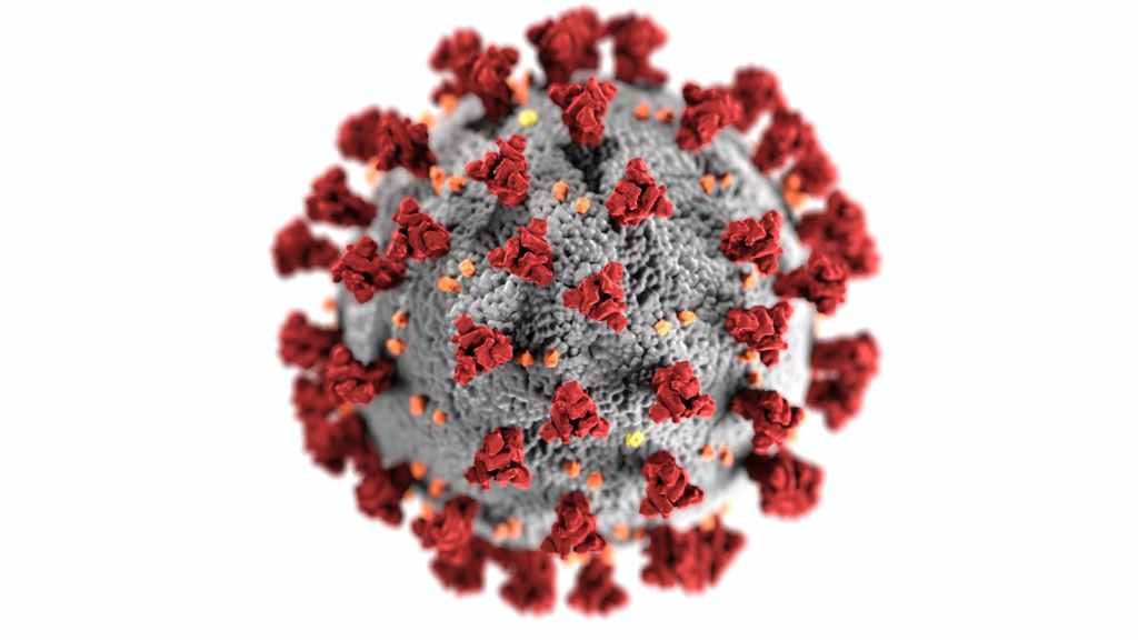
There are many cases of delta and omicron co-infection. When the two variants co-infect a person, an exchange mutation may produce a new viral offspring. The Deltacron strain is a combination of these two.
Professor Leondios Kostrikis, Professor of biological sciences at the University of Cyprus, identified omicron-like genetic signatures within the delta genomes and named it Deltacron.
This deltacron is identified in Cyprus by the Professor and Head of the Laboratory of Biotechnology and Molecular Virology Department.
His team identified 25 Detacron cases. In addition, professor Kostrikis and his team found that this infection is more in hospitalized patients for Covid-19 than in non-hospitalized patients.
According to specialists and scientists, this may be due to laboratory contamination. Deltacron is not absolute and is probably the result of a sequencing error. Experts report no evidence that Omicron and Delta have combined to produce a new variant.
The global health expert Boghuma Kabisen Titanji also reported that it may be due to sample contamination.
Dr.Jeffrey Barrett, Director of the Covid-19 Genomics Initiatives at the Wellcome Sanger Institute, says that ‘this is almost certainly not a recombinant of the Delta and Omicron lineages.
It is suggested that fragments of Omicron have been inserted into Delta’s genetic make-up during the sequencing process when attempting to identify a Cvid variant infection.
Professor Aris Katzourakis, Professor of evolution and genomics at Oxford University, says that it is a standard error in any laboratory.
On seventh January, they sent sequences of the twenty-five deltacron cases to GISAID, the international database that tracks changes in the virus mutation of viruses.
It is unclear whether this deltacron strain exists and, if yes, whether it is more pathological and more contagious than the delta and omicron strains. And it is a matter of concern that deltacron was found more in hospitalized patients with Covid-19 infections than in non-hospitalized Covid-19 cases.
Health Minister of Cyprus said on Sunday, 8, January, that Deltacron isn’t of concern, and later, more details will be given. article”>https://www.bloomberg.com>article
Experts and officials are not concerned by Deltacron.
Professor of virology at the University of Bristol said, ‘It’s not one to worry about.
Florona is a double infection of Covid19 and Influenza viruses. It is the Covid-19-Influenza variant.
The Covid19-Influenza variant is another variation of Coronavirus, like its other variations alpha, beta, gamma, omicron, and Demicron.
Detection of the disease
In Israel, the first case of Florona -a double infection of Covid19-Influenza variant was detected on the last date of 2021, that is 31st December 2021.
This infection was detected in a pregnant woman who came in the hospital -Rabin Medical Center for delivery.
Prevention
It is surprising that in Israel 3 rd dose of covid 19 vaccination is completed. But due to changes in strains of Corona 19 viruses, the infection occurs. Therefore 4 th dose is recommended, especially in aged and immunocompromised persons.
We know that in pregnancy immunity is slightly depressed. Therefore this group must get extra protection.
Israel’s national Health providers started 4 th dose of vaccination against Covid 19 to individuals with compromised immune systems and aged persons.
Measures to prevent Covid 19 disease, as Govt. and WHO guidelines released from time to time, must be followed. Some of them are:
Maintain social distance of 6 feet.
2. Avoid touching anything.
3. Wash your hands frequently with soap and water.
4. Always use masks properly.
5. Avoid social gathering
Link:
Israel -Tel-Aviv on 31 st December Arab news ANI, Al-Arabiya Arab news/mhvr.
This article is to provide knowledge of Purpura, a very common condition the public suffers from. How it is produced, our line of treatment, and much more.
Introduction
Purpura is a clinical condition in which there is a tendency for spontaneous bleeding, usually beneath the skin, mucous membrane, and internal organs. In this condition, bleeding time is prolonged (average is 2 to 6 minutes.), but clotting time is average (standard is five to10 minutes).https://learn-and-fly.co.in/2021/12/26/what-is-bloodcompositionfunctions/(opens in a new tab)
1. Thrombocytopenic purpura-platelet count is 50-10 thousand per cubic milliliter of blood.
2. Thromboasthenic purpura platelet count is average, but the function is abnormal.
Purpura hemorrhagic is severe purpura with bleeding in the skin, mucous membrane, and internal organs and is usually fatal.
Primary purpura is also known as idiopathic, congenital, or hereditary.
I.T.P = Idiopathic thrombocytopenic purpura is due to autoantibodies against platelet membrane glycoprotein IIb-IIa, which causes premature removal of platelets from the reticuloendothelial system.
Allergic purpura is due to some allergies.
Infection by yellow fever, typhus, or infective endocarditis may cause purpura.
Certain drugs, e.g., Iodide, bismuth, ergots, etc., may cause purpura.
Cachectic state: in some cancers, the purpura may occur.
Treatment
1) Small dose of corticoids.
2) Splenectomy.
3) I.V. infusion of immunoglobulins.
4) Platelet transfusion.
Intravenous (I.V.) infusion is a slow therapeutic introduction of fluid other than blood into a vein.
Transfusion, also known as blood transfusion, is the transfer of blood or a blood component, such as plasma or platelets, from one person to another in the vein.
Disclaimer
This article is about academic-health-related topics and not for treatment purposes. If you have any problems, consult a qualified medical professional.


Leave a comment