Table of Contents:
Introduction
The spleen is the largest wedge-shaped, dark purple-colored lymphatic organ of the lymphoid system, and it is connected to the vascular system. It is highly vascular. Its structure is similar to a large lymph node.
Measurements –length-5inches, breadth-3 3 inches,thickness-1 inch. Weight(average) is only 7 ounces.
Spleen performs multiple functions, although it is not essential for survival ,it is vital for healthy life. Dysfunctions of spleen will produce serious problems.
Location
The left hypochondrium houses the spleen between the fundus of the stomach and the diaphragm.
Behind the mid-axillary line opposite the 9th,10th, and 11th ribs.
External features Macroscpic Feature
Two ends, two surfaces, two angles, and three boundaries.
Two ends: (1) anterior or lateral end that faces downward and forward, (2) posterior or medial face that faces upward and backward.
Three boundaries: 1. superior,2.inferior, and 3. intermediate.
The superior border is convex and faces and is in contact with the undersurface of the left side of the diaphragm. This has one or more notches called the splenic notch.
The inferior border is rounded and corresponds to the lower border of the eleventh rib.
The intermediate border is round and separates the gastric impression from the renal impression.
Two surfaces-1. The diaphragmatic surface is smooth, convex, and directed upward, backward, and to the left.
1. Visceral surface is concave and irregular and has four impressions 1. Gastric impression is the most significant impression and is due to the fundus of the stomach. It lies between the superior and intermediate border of the spleen.
Renal impression is due to the left kidney. It is present below and behind the gastric impression and lies between the intermediate and inferior boundaries.
The left colic flexure produces a colic impression. It is triangular and situated in front of the lateral end. and
The tail of the pancreas causes the pancreatic impression, which is located between the hilum and the colic impression.
There is a groove through which the splenic artery and nerves enter, and veins and lymphatic exit.
Relation of the Spleen
The spleen is surrounded by peritoneum and suspended by four ligaments.
1. Gastrosplenic ligament- This ligament extends from the hilum of the spleen to the upper one-third of the greater curvature of the stomach. Short gastric vessels are present in this ligament.
2. Splenorenal ligament- This ligament extends from the hilum of the spleen to the anterior surface of the left kidney. Tail of the pancreas, splenic venules, and pancreatosplenic lymph nodes are present in this ligament.
3. Colicosplenic ligament
4. Phrenocolic ligament
The visceral surface of the spleen is related to the fundus of the stomach, the anterior surface of the left kidney, the left colic flexure, and the tail of the pancreas.
Near the middle of the spleen, a long fissure is present, which is known as the hilum.
Penicilliary radicles are arterioles that supply germinal centers.
Develops from mesenchymal tissue, while most of the gut organs are derived from endoderm.
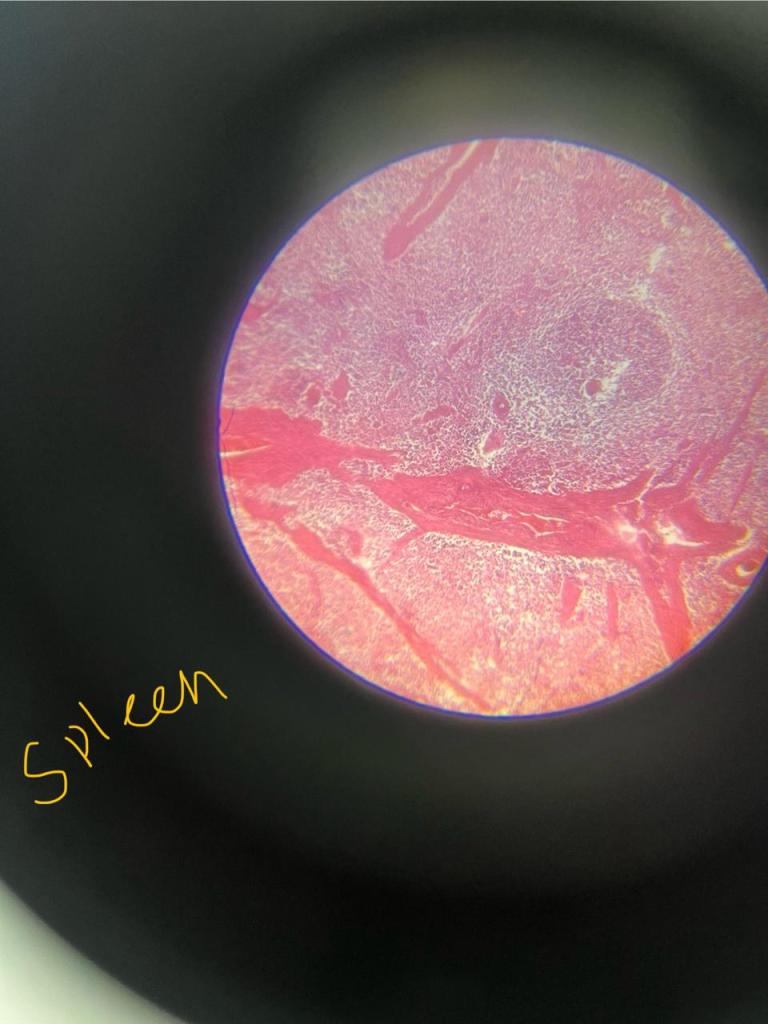
Histology
The outer capsule covers the spleen, made up of thick fibrous tissue.
Beneath the capsule, there is a subcapsular space. The spleen contains two different types of tissues, white pulp and red pulp.
The white pulp is white in colour and contains growing immune and blood cells. White pulp is composed of Malpighian corpuscles. These include lymphoid follicles rich in B lymphocytes and periarteriolar lymphoid sheaths rich in T lymphocytes.
The red pulp is red in colour and contains sinuses filled with blood, reticular fibers, and a marginal zone bordering on the white pulp.
Blood supply
The Splenic artery, the largest branch of the coeliac artery, provides the main blood supply. At the hilum, it divides into five or more branches and enters the spleen. Within the spleen, it divides repeatedly into straight arterioles called Penicilliary radicles. The penicilliary radicles supply the germinal centers.
Blood supply of the Spleen
Close theory: capillaries open directly in the venous sinusoids and lie in the red pulp.
Open theory: capillaries open into red pulp from where blood enters sinusoids through their fenestrated wall.
Compromise theory: according to this theory, circulation is open in the distended spleen and closed in the contracted spleen.
Lymphatic drainage
The Spleen has only efferent lymphatic vessels. There are no lymphatics in the proper splenic tissue. The tissue fluid formed in the spleen freely enters the venous sinusoids. However, the splenic lymphatics are present in the capsule, trabeculae, and visceral peritoneum, and drain into the pancreatocoliecal lymph nodes.
Venous drainage
The splenic vein is formed by the confluence of venules at the hilum, which join the superior mesenteric vein to form the portal vein. Its tributaries are 1. the short gastric vein, 2. the left gastroepiploic vein, 3. the pancreatic vein, and inferior mesenteric vein.
Nerve Supply
Sympathetic fibre derived from the celiac plexus supplies smooth muscles present in the blood vessels, as well as in the trabecular cord.[ splenic plexus, which connects a branch of the celiac ganglion to the vagus nerve.
Functions
1. Filtration of blood
2. Reservoir of blood cells
3. Active immune response: cell-mediated and humoral immunity.
4. Produces opsonins, properdin, and tuftsin.
5. Hematopoiesis in the embryo and in case of emergency.
6. Maintains homeostasis.
Clinical
An enlarged spleen is splenomegaly.
Hyperactive spleen is hypersplenism.

Leave a comment